Journal of Animal Health and Production
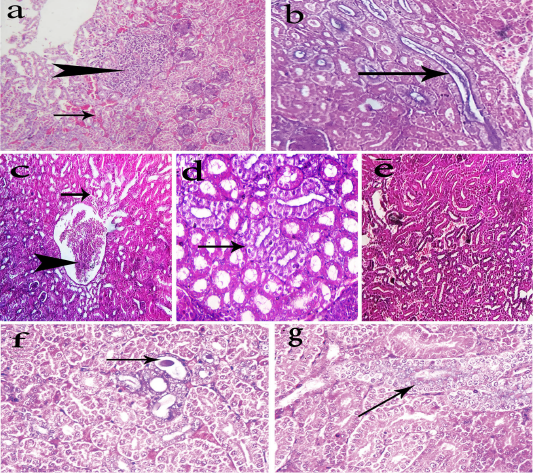
Photomicrographs of H&E stained kidney sections of the chickens. The kidneys of group-1 shows intertubular extravasated RBCS (arrow) with focal necrosis replaced by leukocytes cells (head arrow) (a, 200x), and urate deposition (arrow) within renal tubules (b, 400x). The kidneys of group-2 shows mild tubular degeneration (arrow) and congestion of the renal tubules (head arrow) (c, 200x), nephrotic changes in some renal tubules (arrow) (d, 400x), and few extravasated erythrocytes among dissociated renal tubule (e, 200x). The kidneys of group-3 shows dilatation of some renal tubules containing casts (arrow) (f, 400x) and regeneration of some renal tubules (arrow) (g, 400x).
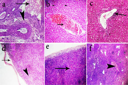
Photomicrographs of H&E stained liver and spleen sections of the chickens. Livers of group-1 shows portal fibrosis (head arrow) and cholestasis(arrow)(a, 400x). Livers of group-2 shows moderate congestion of the hepatic blood vessels (arrow) (b, 200x). Livers group-3 shows mild perivascular lymphocytic cells infiltration of (arrow) (c, 200x). Spleen of group-1 shows lymphoid depletion (head arrow) and splitting of capsule (arrow) (d, 400x). Spleen of group-2 shows subcapsular edema and mild depletion (arrow) (e, 200x). Spleen of group-3 shows congestion of blood vessels (arrow head) with hyperplastic white pulps (f, 200x).
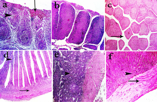
Photomicrographs of H&E stained bursa of fabricius and intestine sections of the chickens. Bursa of fabricius of group-1 shows mild depletion of lymphocytes (head arrow), interfollicular fibroplasia and focal hemorrhage (arrow) (a, 400x). Bursa of fabricius of group-2 shows apparently normal lymphoid follicles (b, 200x). Bursa of fabricius of group-3 shows mild interfollicular edema (arrow) (c, 200x). Intestine of group-1 shows degenerated submucosal glands (arrow) and fusion of villous tips (head arrow) (d, 400x). Intestine of group-2 shows hyperplastic intestinal glands in submucosa (head arrow) (e, 400x). Intestine of group-2 shows hyaline degeneration of muscular layer (arrow) and proliferative submucosal glands (f, 400x)


