Journal of Animal Health and Production
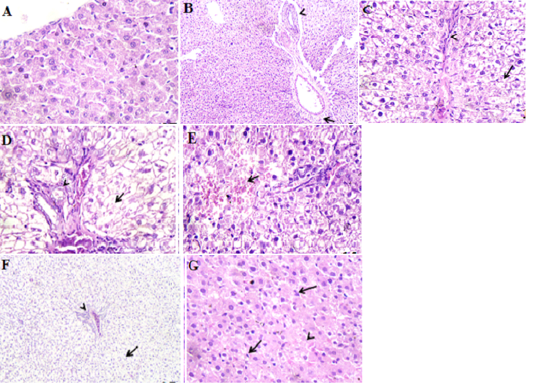
Photomicrographs of rat liver. A. section of control group showing normal hepatic parenchyma. H&E X400. B. etoricoxib group (on day 14 post administration) showing hydropic degeneration, edema of portal vein (arrow) and periductular fibrosis (arrow head). H&E X120. C. azithromycin group (on day 7 post treatment) showing diffuse acute cell swelling or necrosis (arrow) of the hepatic cells beside fibroblasts proliferation in interlobular tissue (arrow head). H&E X 400. D. etoricoxib+azithromycin group (on day 7 post treatment) showing extensive necrosis of the hepatic cells (arrow) and numerous bile ductules (arrow head) in the portal area. H&E X400. E. etoricoxib+azithromycin group (on day 14 post treatment) showing necrotic hepatic cells replaced by extravasted erythrocyte (arrow). H&E X400. F. etoricoxib+azithromycin group received ascorbic acid (on day 7 post treatment) showing mild reversible change (cloudy swelling) of the hepatic cell (arrow) and hyperemic portal vein (arrow head). H&E X120. G. etoricoxib+azithromycin group received ascorbic acid (on day 14 post treatment) showing vacuolar degeneration of the hepatic cells (arrow) and hyperplastic kupffer, s cells (arrow head). H&E X400.


