Journal of Animal Health and Production
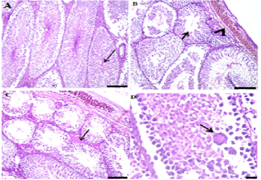
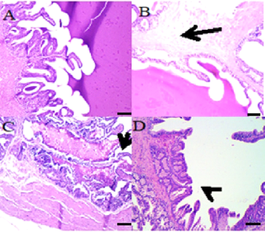
A: Photo micrograph of male rat testis (control) showing normal semineferous tubules and active spermatogenesis and the testicular seminiferous tubules and covering tunica albuginea appeared normal. (H&E; x 100). B: Photo micrograph of male rat Testis reveals moderate spermatogenesis (arrowhead) of congested blood vessels in tunica albuginea, The majority of seminiferous tubules showed moderate spermatogenesis and congested blood vessels and in interstitium and tunica albuginea (H&E; x100). C: Photo micrograph of male rat Testis showing mild spermatogenesis and degenerated spermatocytes (thin arrow) with congested blood vessels (thick arrow), some seminiferous tubules exhibited mild spermatogenesis besides edema and congested blood vessels (H&E x100). D: Photo micrograph of male rat testis showing intense degenerated necrotic spermatocytes (thin arrow) with spermatid giant cells (thick arrows), the majority of tubules had vacuolated and degenerated or necrotized spermatogonial and spermatocytes beside spermatid giant cells (H&E; x400).
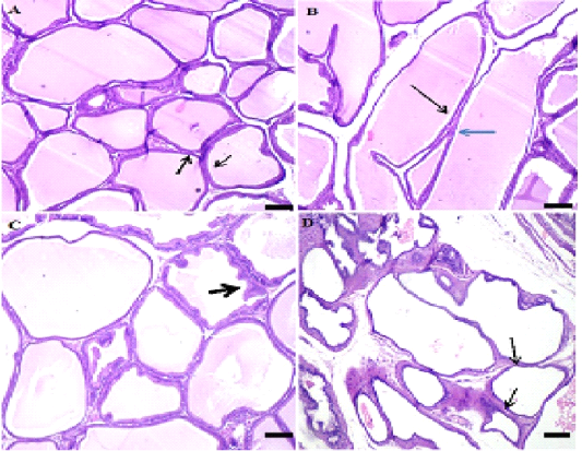
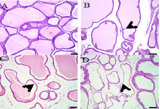
A: Photo micrograph of male rat seminal vesicle (control) showing normal tubule-alveolar glands and secretion, trabeculae and covering Tunica were normal (H&E x100). B: Photo micrograph of male rat seminal vesicle showing distended glands with secretion and distended ducts and glands with secretion with mild atrophy of some epithelial lining of a few glands (H&E; x100) . C: Photo micrograph of male rat Seminal vesicles showing hyperplastic tubuloalveolar glands (arrows) containing little secretion, normal tubule alveolar glands and some were hyperplastic and contain moderate secretion (double arrow) (H&E; x100). D: Photo micrograph of male rat Seminal vesicle showing atrophied tubular glands with retained secretion (arrows), a few tubuloalveolar glands lined by atrophied epithelium with intense secretion in its Lumina (H&E; x100).
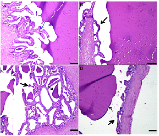
A: Photo micrograph of male rat Prostate (control) one day post end of administrated extract of nasturatium flower showing normal prostatic acini with secretion and the glandular tissue and stroma were normal (H&E; x100). B: Photo micrograph of male rat prostate gland (Flower I) Showing apparently normal acini containing secretion (both black and blue), all the glandular tissue contains secretion and were normal (H&E; x100). C: Photomicrograph of male rat prostate gland (Flower II) showing apparently normal acini containing little secretion (arrows), all glandular tissue appeared normal and contain a little secretion (H&E; x100). D: Photo micrograph of male rat Prostate (Flower III) showing cystic dilated prostatic acini devoid from secretion (arrow), some acini appeared cystic and lined by flattened epithelium and devoid from secretion (H&E; x100)
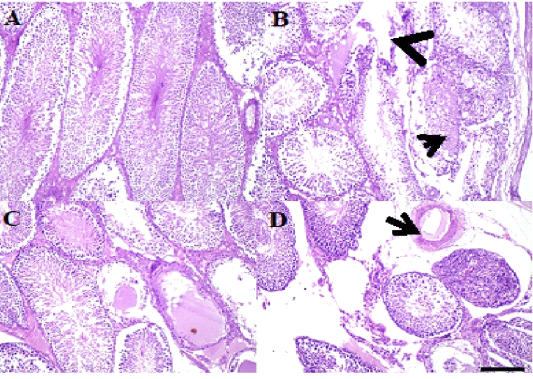
A: Photo micro graph of male rats (control) showing normal seminiferous tubules and active spermatogenesis, seminiferous tubules, interstitium and covering were within normal morphological picture (H&E; x100). B: Photo micro graph of male rat testis showing necrotic seminiferous tubules containing edema and necrotic debris (arrow) and interstitial edema (arrowhead), some seminiferous tubules were necrotic and contained edema and necrotic debris beside interstitial edema, A few tubules had pyknotic spermatogonia and spermatocytes with intense congestion and edema in the interstitium (H&E; x100). C: Photo micro graph of male rat showing intense edema and necrosis of the seminiferous tubules and arrest of spermatogenesis (arrows), the majority of tubules showed arrest of spermatogenesis with intense necrosis and edema inside their lumina severe edema and congested blood vessels in capsule and interstitium (H&E; x100). D: Photo micro graph of male rat testis showing spindle cell invasion for necrotic seminiferous tubules (arrows) and interstitial edema, the majority of necrotic acini invaded by spindle cells with mononuclear cells infiltration beside interstitial edema , other tubules collapsed with loss of thus lining epithelium and contained necrotic debris and threads. Dilated blood vessels with prominent edema in interstitium (arrow) (H&E; x100).
A: Photo micro graph of male rat seminal vesicle (control) showing normal tubuloalveolar glands and secretion, glandular structure, Trabeculae and covering tunica were normal (H&E; x100) . B: Photo micro graph of male rat Seminal vesicle showing normal tubule alveolar glands and edema in the smooth muscles, all the glandular structure appeared normal while tunica muscularis were edematous arrows (H&E; x100). C: Photo micro graph of male rat seminal vesicle showing partial desquamation of some glandular epithelium (arrows) and foamy secretion, some glands had partially desquamated epithelium and filled with foamy esinophilic colloidal secretion (H&E; x100). D: Photo micro graph of male rat seminal vesicle showing hyperplastic glands without secretion (arrows), tubuloalveolar glands were either hype plastic without secretion. Tunica muscularis had partial edema or necrosis (H&E; x100).
A: Photo micro graph of male rat Prostate gland (control) showing normal prostatic acini with secretion, the acini, interstitium and muscles were normal (H&E; x100). B: Photo micro graph of male rat prostate gland showing normal prostatic acini (arrows) and interstitial edema, the majority of prostatic acini contained secretion and normal beside interstitial edema (H&E; x100). C: Photo micro graph of male rat prostate gland showing slightly cystic acini with little secretion (arrows), mild dilated acini contained little secretion beside edema and congested blood vessels in interstitium (H&E; x100). D: Photo micro graph of male rat prostate gland showing cystic acini without secretion (arrows) and interstitial edema, Cystic glandular acini devoid from their secretion and interstitial edema were common, other acini lined by hyperplastic epithelium (H&E; x100).


