Journal of Animal Health and Production
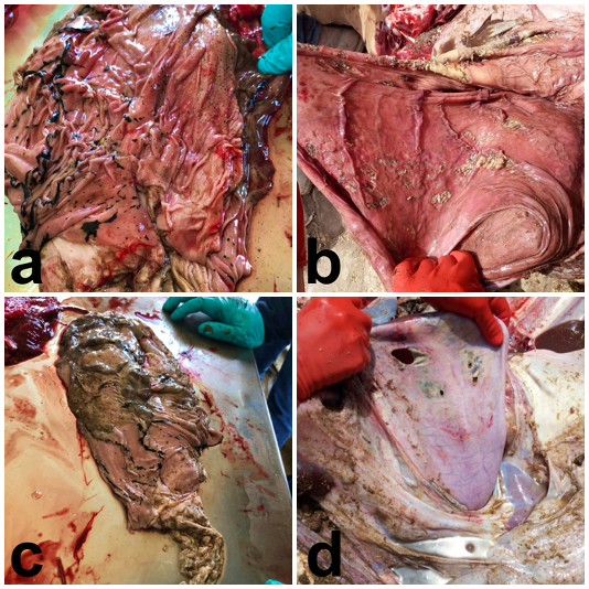
Calves, abomasa, different degrees of lesion severity. (a) several ulcers in fundus are evident, some lineal on the folia and other irregulars with blood clots on the surface. Microscopically, few were transmural and reached the serosa provoking focal peritonitis but not rupture. These lesions could be included as grade 2 (Marshall, 2009). (b), other ulcers were covered by fibrinous exudate (diphtheric plaque); histologically these lesions reached muscular layer wall but not the serosa (grade 1). (c) In some cases, the grade 2 ulcerative abomasitis included a profuse haemorrhage forming large blood clots within the abomasum. In this case the clot is on the upper left corner. (d) In some necropsies the abomasal content was spread throughout the peritoneal cavity leading to mild and diffuse fibrinous inflammation within the abdominal cavity (grade 4; focal peritonitis is included as grade 3). In all cases mild to severe bronchopneumonia was recognized (grossly and microscopically), except in most cases of grade 4, abomasal perforating ulcers, in which bronchopneumonia lesions were inconclusive, because limited extension (≤ 30%).
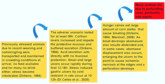
Proposed pathogenesis of the condition described.


