Journal of Animal Health and Production
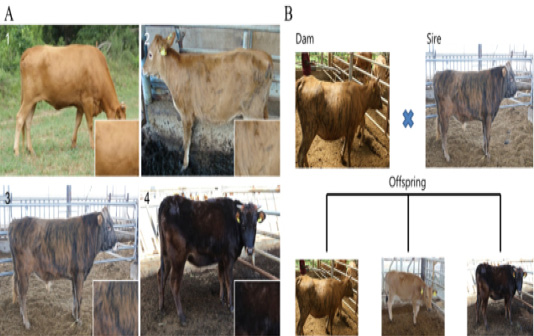
Different patterns of expression of coat color between Korean native cattle (Kim et al., 2013). A: Differences in the cattle coat color determined by the ratio of Yellow: Black. 1) Hanwoo (10:0), 2) Korean brindle cattle (Brindle coat color type 1 (9:1)), 3) Korean brindle cattle (Brindle coat color type 2 (4:6)). B: The difference in the coat color of the offsprings was evaluated based on the crossbreeding of the cattle.
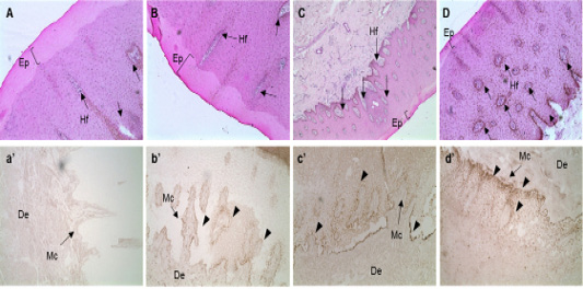
In situ hybridization of MC1R mRNA in the follicular zone of cattle. Black bar = 100 μm. Black arrows indicate MAP1LC3A RNA probe detection. MC1R probe was made using NCBI genetic information EU169234, sequence is “ctgtgtctga cttgctggtg agcgtcagca acgtgctgga gacggcagtc atgccgctgc tggaggccgg tgtcctggcc acccaggcgg ccgtggtgca gcagctggac aatgtcatcg”. A, B, C and D: H&E staining of skin tissues of cattle. a’, b’, c’ and d’: in situ hybridization of MC1R probe detection dots. *, ** Different letters within the same column represent a significant difference (p<0.05). A-a’) Hanwoo (HC), B-b’) Korean brindle cattle, C-c’) Korean dark cattle, D-d’) Holstein cattle. Hf: Hair follicle, Ep: epidermis, Mc: Melanocyte zone, De: dermis. A–D: 100X magnification; a’-d’: 200X magnification.
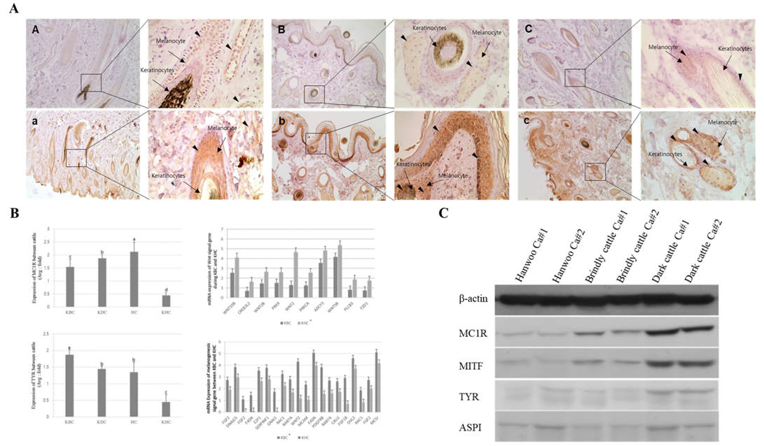
Expression analysis of melanogenesis-associated genes between the Korean native cattle. A: Immunodetection of MC1R and TYR protein A-D) Detection of MC1R protein, a-d) Detection of TYR protein, A-a) Hanwoo, B-b) Korean brindle cattle, C-c) Korean dark cattle. B: Real-time qPCR analysis of melanogenesis-associated genes, where KBC) Korean brindle cattle, KDC) Korean dark cattle, HC) Holstein cattle, KHC) Korean Hanwoo cattle. The bars represent the average fold changes in 3 independent experiments (±SD). C: Western blot analysis.


