Journal of Animal Health and Production
Research Article

Retrospective Echocardiographic Survey of Canine Cardiac Conditions in Kuala Lumpur, Malaysia
Yap Kai Sin1, Shanthi Ghanabadi1, Ibrahim Abdul-Azeez Okene1,2*, Amilan Sivagurunathan3, Sivan Ayarhany3
1Faculty of Veterinary Medicine Universiti Malaysia Kelantan, 16100 Kota Bharu, Kelantan, Malaysia; 2Department of Veterinary Surgery and Radiology Faculty of Veterinary Medicine, University of Maiduguri 600244 Maiduguri, Nigeria; 3Animal Medical Centre Sdn Bhd, Wisma Medivet, 8, Jln Tun Razak,50400 Kuala Lumpur, Malaysia.
Abstract | Canine cardiac diseases are routinely diagnosed in veterinary practices across Malaysia. The prevalence and distribution of cardiac diseases in dogs are yet to be documented despite the availability of expertise and imaging modalities in Malaysia’s various small animal practices. This study was conducted to determine the prevalence and distribution of cardiac diseases in dogs in Malaysia using a representative clinic with large caseloads. A three-year retrospective study (2015-2017) was conducted using patient records of 264 dogs from Animal Medical Centre (AMC), Kuala Lumpur. Data involving cardiac conditions and their diagnosis were retrieved and analyzed. Data were presented descriptively in percentages and relationships analyzed using chi-square analysis. Myxomatous mitral valve disease (MMVD) accounted for 65.68% of the cardiac conditions affecting dogs and was the most diagnosed condition using Doppler ultrasonography. Generally, age and breed were strongly associated (p<0.001) with cardiac diseases in dogs. Age and breed play important roles in the occurrence of cardiac diseases in dogs in Malaysia. As the dogs grew older, they tended to show clinical signs (p<0.001) of cardiac diseases. Therefore, adequate precautionary measures, including regular check-ups, strategic exercise, and the dietary regime, are needed to detect cardiac diseases, especially in geriatric dogs.
Keywords | Cardiovascular disease, Dilated cardiomyopathy, Dog, Echocardiography, Myxomatous mitral valve disease.
Received | January 25, 2021; Accepted | January 28, 2021; Published | April 25, 2021
*Correspondence | Ibrahim AAO. Faculty of Veterinary Medicine Universiti Malaysia Kelantan, 16100 Kota Bharu, Kelantan, Malaysia; Email: ibrahim.az@umk.edu.my
Citation | Sin YK, Ghanabadi S, Okene IAA, Sivagurunathan A, Ayarhany S (2021). Retrospective echocardiographic survey of canine cardiac conditions in Kuala Lumpur, Malaysia. J. Anim. Health Prod. 9(2): 198-204.
DOI | http://dx.doi.org/10.17582/journal.jahp/2021/9.2.198.204
ISSN | 2308-2801
Copyright © 2021 Okene et al. This is an open access article distributed under the Creative Commons Attribution License, which permits unrestricted use, distribution, and reproduction in any medium, provided the original work is properly cited.
INTRODUCTION
Imaging modalities are used in medical and veterinary practices to obtain information on clinical conditions that are uneasily diagnosed by other means (Atkins et al., 2007). Laboratory tests and imaging techniques are simultaneously used for the diagnosis of heart disease and heart failure in the medical field (Atkins et al., 2009) and small animal cardiology (Bonagura et al., 1985). Imaging techniques provide a non-invasive, closely accurate, accessible, cheap, and convenient means for diagnosing cardiac conditions with minimal discomfort to the patient (Boon, 2011).
Among the diagnostic imaging modalities for investigating cardiac diseases, echocardiography is the most common and useful (Borgarelli et al., 2008). Echocardiography enables the veterinarian to diagnose abnormal presentation of heart by determining the size and cardiac silhouette (Boon, 2011). The images may also provide clues to the specific disease present (Borgarelli and Haggstron, 2010). Echocardiography images are captured in real-time and it reveals the structure and movement of the internal organs (such as the heart) including the blood flow (Brown et al., 2007). Veterinarians prefer echocardiography for cardiac examination as it is non-invasive, able to gives clear images of internal soft tissues, and presents minimal hazards to both the clinician and patient (Chetboul and Tissier, 2012).
For over three decades, echocardiography has been the most critical and cost-effective diagnostic imaging modality in clinical cardiology (Atkins et al., 2009; McGregor, 2014). Echocardiography is a diagnostic method, which allows non-invasive and non-ionizing visualization of structures and components of the heart using ultrasound. Such components include the atria and ventricles, aorta, auricular appendages, and all the valves (Chetboul and Tissier, 2012). Doppler echocardiography is used on a specific location to provide information on blood flow when the sound waves pass through the heart and vessels (Rantanen and Ewing, 2005).
The use of echocardiography has contributed greatly to veterinary cardiology in diagnosing various cardiac abnormalities such as myxomatous mitral valve disease, dilated or hypertrophic cardiomyopathies and cardiac shunts. It has enabled early interventions and improved the prognosis of management resulting directly from prompt diagnosis. Apart from this, cardiac abnormalities can be detected by using echocardiography which includes mitral valve regurgitation, tricuspid valve regurgitation, valvular lesions, pleural and pericardial effusions, myocardial diseases, cardia shunts, stenotic lesions, and patent ductus arteriosus (King, 2006). The status and severity of cardiac conditions can be assessed through echocardiography by determining the cardiac chamber size, normal function of heart, the speed of the blood flow and hemodynamic of heart (Slupe et al., 2008). Globally, the incidence of heart diseases in dogs is estimated at 10 – 15% of general cases (MacPete, 2018). In Malaysia the prevalence and classification of cardiac diseases has received little attention despite widespread usage of echocardiography in most small animal clinics. Nevertheless, echocardiographic examination of the heart opened a new dimension in the understanding and classification of heart conditions in dogs. This study was conducted to determine the prevalence and distribution of cardiac conditions in dogs in Kuala Lumpur, Malaysia.
MATERIALS AND METHODS
Ethical approval
This retrospective study was approved by the Faculty Animal Care and Use Committee, Faculty of Veterinary Medicine, Universiti Malaysia Kelantan with an approval number is UMK/FPV/FYP/2018/20.
Study design
This study retrospectively assessed 3 years echocardiographic records of 264 dogs managed at the Animal Medical Centre, Kuala Lumpur between January 2015, and December 2017. Presence of clinical signs of cardiorespiratory disease including cough, dyspnea, exercise intolerance and syncope were the inclusion criteria used. Prevalence and distribution of cardiac conditions were determined. Data were classified according to age, breed, and sex of patients as obtained from the patient records. Breeds include Shih Tzu, Bichon fries, Jack Russell, Pomeranian, Daschund, Poodle, Cavalier King Charles Spaniel, Silky terrier, Chi huahua, Mini Pinscher, Pekingese, Maltese, Pug and Yorkshire Terrier. Percentage of cardiac conditions among the dogs presented was calculated. Age was categorized into two groups viz; < 7 years old and > 7 years old. Breeds were categorized as small, medium, and large breeds.
Echocardiography technique
Routine examinations were conducted with dogs placed on either right or left lateral recumbency. Right parasternal long axis four-chamber view (RPSL), right parasternal short axis (RPSA) papillary muscle level, right parasternal short axis mitral valve level and right parasternal short axis left atrium/aorta were evaluated on right lateral recumbency. M-mode echocardiography was evaluated at right ventricular papillary muscle level, mitral valve level, and left atrium/aorta level. Left apical four chamber was evaluated on left recumbency. Doppler echocardiography was used to determine blood flow from the atrium to ventricle.
Statistical analysis
Obtained data were expressed descriptively using percentages for prevalence of cardiac conditions while Chi-square test was used to determine the association of cardiac abnormalities with age, breed, and sex. Findings were considered signification at p<0.05.
RESULTS
Prevalence of cardiac conditions in dogs
Among 264 of dogs involves in this study, a total of 206 dogs were diagnosed of cardiac conditions at the AMC between January 2015 and December 2017. Annual prevalence observed showed that in 2015, 66.23% of dogs had MMVD, 6.49% had dilated cardiomyopathy (DCM), while 9.10% were diagnosed with other heart diseases as shown in Figure 1. While the remaining 18.18% showed no evidence of heart diseases even though signs were present, but more of respiratory diseases. In 2016, MMVD was the highest diagnosed heart diseases, which accounted for 66.30% of cases, followed by 9.78% of dogs diagnosed with other heart diseases and 3.26% with DCM, while the remaining 20.66% had no evidence of heart disease. For 2017, 64.21% of dogs presented were diagnosed with MMVD, 6.32% with DCM, and 3.15% with other heart diseases while 26.32% showed no evidence of heart disease.
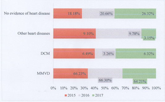
Figure 1: Common canine cardiac conditions diagnosed at Animal Medical Centre, Kuala Lumpur from 2015 to 2017.
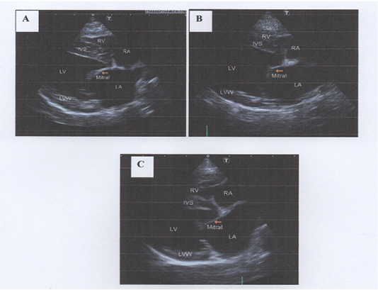
Figure 2: Thickened mitral valve at right parasternal long axis four-chamber view. 2-A: Mild thickening of mitral valve, 2-B: Moderate thickening of mitral valve and prolapse of the anterior leaflet, 2-C: Severe thickening of mitral valve and mitral valve prolapse (MVP) of the anterior leaflet towards left atrium.
Myxomatous mitral valve degeneration is the most common cardiac condition diagnosed at the Animal Medical Centre, Kuala Lumpur. The characteristics of MMVD diagnosed using echocardiography were (Figures 2, 3 and 4). Thickened mitral valve seen on right parasternal long axis four-chamber view. Mild (Figure 2-A), moderately thickened mitral valve and prolapse of the anterior leaflet (Figure 2-B) and severely thickened mitral valve and prolapse of the anterior leaflet (Figure 2-C). The degree of mitral regurgitation into left atrium was obtained at right parasternal long axis four-chamber view using Doppler echocardiography (Figure 3). Mild (Figure 3-A), moderate (Figure 3-B) and severe mitral regurgitation (Figure 3-C) were observed. Dilation of left atrium was also seen on right parasternal short axis left atrium / aorta view and classified as mild (Figure 3A), moderate (Figure 3-B) and severe dilation of left atrium (Figure 3-C).
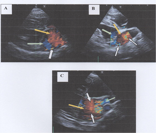
Figure 3: Degree of mitral regurgitation (MR) into left atrium at right parasternal long axis four chamber view using Doppler echocardiography. In 3-A, there is mild mitral regurgitation in to left atrium with mild baseline colour scale is shifted and presence of less than 10% of colour aliasing. In 3-B, there is moderate mitral regurgitation with moderate baseline colour is shifted and presence of 50% of colour aliasing. In 3-C, there is severe mitral regurgitation with severe baseline colour scale is shifted and presence of more than 50% of colour aliasing. Note: Yellow arrows show shades of red indicates the bloods flow towards transducer; Green arrows show shades of blue indicates the blood flow directed away from transducer; White arrows show the presence of colour aliasing).
Prevalence of canine cardiac conditions in Kuala Lumpur in relation to age, breed, sex and clinical signs
Table 1 shows the prevalence of cardiac conditions according to age, breed, sex and findings during physical examination. Dogs aged 7 years and above (96%) were more consistently diagnosed with MMVD in the three-year study period as compared to younger dogs (< 7 years) accounting for 4.0% prevalence. In the same manner, 100% of dogs diagnosed with DCM were more than 7 years old. Dogs diagnosed of other heart diseases had 68.40% older than 7 years old, while 31.60% were less than 7 years old.
Small breed dogs presented at AMC from January 2015 to December 2017 had the highest prevalence of which 67.60% were diagnosed with MMVD. Medium and large breed dogs had 30.60% and 1.70% respectively for MMVD
Table 1: Descriptive analysis with correlations for dogs diagnosed with cardiac conditions at the AMC, Kuala Lumpur from 2015 to 2017
|
Variable |
Number of dogs (%) |
p-value |
||||
| MMVD | DCM | Other heart diseases | ||||
|
Age < 7 years old > 7 years old |
7 (4.00) 166 (96.00) |
0 (0.00) 14 (100.00) |
6 (31.60) 13 (68.40) |
<0.001 |
||
|
Breed Small Medium Large |
117 (67.60) 53 (30.60) 3 (1.70) |
1 (7.10) 5 (35.70) 8 (57.10) |
11 (57.90) 8 (42.10) 0 (0.00) |
<0.001 |
||
|
Sex Male Female |
94 (54.30) 79 (45.70) |
7 (50.00) 7 (50.00) |
10 (52.60) 9 (47.40) |
0.878 |
||
|
Clinical signs Absent Present |
102 (59.00) 71 (41.00) |
8 (57.10) 6 (42.90) |
6 (31.60) 13 (68.40) |
|||
AMC = Animal Medical Centre, MMVD = Myxomatous mitral valve disease, DCM = Dilated cardiomyopathy.
Furthermore, large breed dogs were the most frequently diagnosed with DCM (57.10%) followed by medium (35.7%) and small (7.10%) breed dogs.
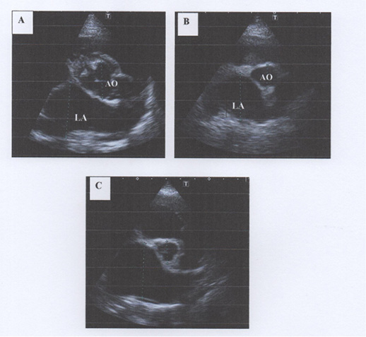
Figure 4: Dilation of left atrium (LA) on right parasternal short axis left atrium/aorta (AO) view. A: Mild dilation of left atrium with LA to Aorta ratio of 1.56, B: Moderate dilation of the left atrium with LA:Ao of 2.04 and C: Severe dilation of left atrium with LA:Ao of 2.47. Note that the normal LA:Ao ratio should be <1.3 (Reproduced from Rishniw et al., 2000)
Data also showed that 54.30% of dogs diagnosed MMVD were male and the remaining 45.7% were female. As for dogs with DCM, both male and female showed an equal distribution (50.0%), while dogs diagnosed with other heart diseases showed 52.60% of male and 47.40% of female.
Clinical signs of MMVD was observed in 41% of dogs from January 2015 to 2017 while 59% were asymptomatic. Forty-three percent of dogs diagnosed with DCM showed clinical signs during routine physical examination while 58% were asymptomatic. Clinical signs for other heart diseases were observed in 68.40% of dogs during routine physical examination while 31.60% were asymptomatic. There was however a significant (p<0.001) association of clinical signs with diagnosis of cardiac disease.
Obtained data revealed a significant statistical difference between the age, breed and cardiac diseases (p<0.001) but no significant statistical difference between sex and cardiac diseases (p=0.878).
DISCUSSIONS
The overall prevalence of the most diagnosed heart condition in dogs MMVD was 65.58% over the three-year retrospective study period. This prevalence of MMVD in the AMC Kuala Lumpur is closely like other reports showing that MMVD account for 70% of all cardiac conditions of dogs (Atkin et al., 2009; Parker et al., 2006). While this study focused solely on the use of echocardiography for diagnostic confirmation, the previous studies included other diagnostic tools including auscultation and routine ultrasonography. Inclusion of these other diagnostic tools accounted for the 4.42% difference in cases prevalence between our findings.
Risk factors including age, breed, and sex were included in this study to ascertain the epidemiology of cardiac conditions in dogs in the sample population. There was a significantly higher association of cardiac diseases with age (p<0.05) where dogs above 7 years had higher prevalence particularly of MMVD (Mohammed et al., 2010). In the same vein, small dog breeds presented at Animal Medical Centre, Kuala Lumpur had a higher incidence of MMVD compare to medium and large breed dogs. Large breed dogs had a higher incidence of DCM compare to medium and small breed dogs. There was a strong relationship between dog breeds and the occurrence of cardiac conditions (p<0.05). It is believed that mutation of linked gene, insulin-like growth factor (IGF-1) carried by small breed dogs increase the susceptibility of MMVD compare to large breed dogs (Jones et al., 2008; Parker et al., 2006). Besides, ancestors of small breed dogs (such as Shih Tzu, Pekingese) that carry SNP haplotype and contribute the inherited genome to the next generation prone to develop MMVD (Palermo et al., 2011).
Furthermore, large breed dogs were more prone to develop DCM compared to small breed dogs in this study. There is number of studies showed that development of DCM is related to genes (Janus et al., 2015; Mattin et al., 2015). It is an acquired heart failure observed most in large and giant breed dogs (Lobo et al., 2010; Swift et al., 2017). Genes associated with canine DCM are dystrophin gene (DMD) in German short-haired pointers, pyruvate dehydrogenase kinase 4 gene (PDK4) in Doberman Pinschers, and striatin gene (STRN) in Boxers, in addition to a locus on chromosome 5 in Doberman Pinschers are believed to lead to the development of DCM (Martin et al., 2010; Orvalho, 2017).
Ninety-six percent of dogs more than 7 years old were diagnosed with MMVD. Besides, small breed is the most common breed diagnosed with MMVD in Animal Medical Centre. Males showed a higher prevalence of MMVD compared to females, 54.30%, and 45.70% respectively. This statement supported the study by Atkins et al. (2009) showing that MMVD is approximately 1.5 times more common in males than in females. In addition, Animal Medical Centre implemented the geriatric program for dogs more than 7 years old for the annual check-up and pre-surgical screening to determine any underlying disease prior to surgery. Fifty-nine percent of dogs diagnosed with MMVD did not show any clinical signs while the remaining 41.00% (Table 1) showed clinical signs upon physical examination. This is consistent with a study done on canine degenerative myxomatous mitral valve disease (Borgarelli et al, 2008; Borgarelli and Haggstrom, 2010). The prevalence of MMVD is associated with age and breed (Maleki and Esmaeilzadeh, 2012).
The echocardiographic findings of MMVD showed thickened mitral valve at right parasternal long axis view, mitral regurgitation into left atrium at right parasternal long axis view using Doppler echocardiography. Mitral valve prolapse is one of the echocardiographic findings in MMVD. Dilation of left atrium was evaluated at right parasternal short axis left atrium / aorta view. This is in consistent with a study by Borgarelli and Haggstrom (2010) which stated prolapse or thickening of one or both mitral valve leaflets is the echocardiographic characteristics of MMVD. Echocardiography provides important information on the severity of disease, such as dilation of left atrium, fractional shortening, LA/Ao ratio, mitral regurgitation during systole and diastole (Borgarelli and Haggstrom, 2010). The recently published American College of Veterinary Internal Medicine (ACVIM) consensus statement recommends that echocardiography should be performed to answer specific questions regarding the cause of the murmur of mitral regurgitation and presence of cardiac chamber enlargement in dogs with suspected MMVD (Atkin et al., 2007; Atkins et al., 2009; Spalla et al., 2016).
Dogs with heart disease should have a proper management regime to maintain their quality of life thus preventing further progression to advanced stages (Atkins et al., 2009; Gugjoo et al., 2014). Asymptomatic heart disease dog that has been diagnosed with heart disease should avoid exercise exhaustion to prevent further exaggerated condition (Parker and Kilroy-Glynn, 2012). Besides, dogs with mild congestive heart failure, repetitive activities and exercise should be avoided (Atkins et al., 2009). Additionally, dogs with severe congestive heart failure which intervention of emergency treatment require hospitalization and should be strictly limited to cage rest only (Atkins et al., 2007).
Medical treatments are provided based on the clinical signs of congestive heart failure, heart murmur grading, radiographic and echocardiography findings of heart disease dogs (Reynolds et al., 2012). Dogs with acute heart disease require hospitalization and medical intervention to control the underlying conditions (Atkins et al., 2009). Loop diuretic, vasodilator, aldosterone antagonist and inodilator were the common drugs used for controlling underlying clinical signs based on the prognosis of the disease for dogs that treat as an outpatient (Borgarelli et al., 2008; Lombard et al., 2006; Parker-Kilroy-Glynn, 2012).
CONCLUSIONS
Myxomatous mitral valve degeneration is the most common canine cardiac conditions diagnosed at Animal Medical Centre, Kuala Lumpur from 2015 to 2017 with a 65.68% prevalence. Age and breed were the risk factors significantly associated with the occurrence of cardiac diseases in dogs in Malaysia. Geriatric dogs above 7 years were the most highly diagnosed with cardiac diseases including MMVD and DCM. Small dog breeds were mostly diagnosed with MMVD in this study while large breed dogs commonly had DCM.
CONFLICT OF INTERESTS
The authors declare that they have no conflict of interest.
AUTHORS’ CONTRIBUTION
YKS, SG and IAO designed the study, interpreted the data, and drafted the manuscript. YKS, AS, and SA were involved in collection of data and contributed in manuscript preparation. IAO, SG and YKS took part in preparing and critical checking of this manuscript.
REFERENCES


