Journal of Animal Health and Production
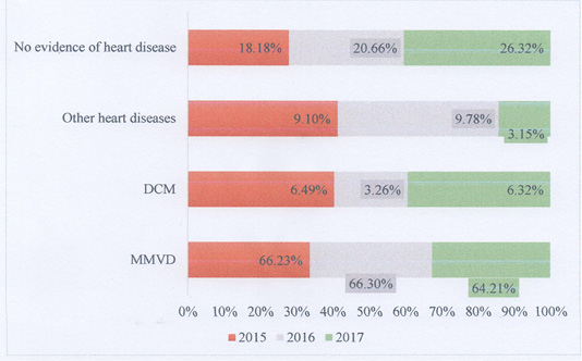
Common canine cardiac conditions diagnosed at Animal Medical Centre, Kuala Lumpur from 2015 to 2017.
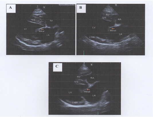
Thickened mitral valve at right parasternal long axis four-chamber view. 2-A: Mild thickening of mitral valve, 2-B: Moderate thickening of mitral valve and prolapse of the anterior leaflet, 2-C: Severe thickening of mitral valve and mitral valve prolapse (MVP) of the anterior leaflet towards left atrium.
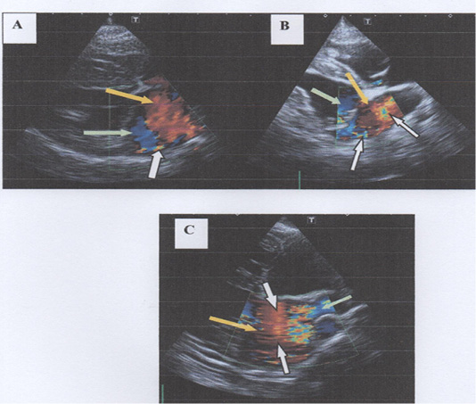
Degree of mitral regurgitation (MR) into left atrium at right parasternal long axis four chamber view using Doppler echocardiography. In 3-A, there is mild mitral regurgitation in to left atrium with mild baseline colour scale is shifted and presence of less than 10% of colour aliasing. In 3-B, there is moderate mitral regurgitation with moderate baseline colour is shifted and presence of 50% of colour aliasing. In 3-C, there is severe mitral regurgitation with severe baseline colour scale is shifted and presence of more than 50% of colour aliasing. Note: Yellow arrows show shades of red indicates the bloods flow towards transducer; Green arrows show shades of blue indicates the blood flow directed away from transducer; White arrows show the presence of colour aliasing).
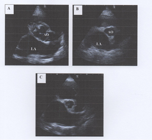
Dilation of left atrium (LA) on right parasternal short axis left atrium/aorta (AO) view. A: Mild dilation of left atrium with LA to Aorta ratio of 1.56, B: Moderate dilation of the left atrium with LA:Ao of 2.04 and C: Severe dilation of left atrium with LA:Ao of 2.47. Note that the normal LA:Ao ratio should be <1.3 (Reproduced from Rishniw et al., 2000)


