Journal of Animal Health and Production
Case Report
J. Anim. Health Prod. 9(2): 136-139
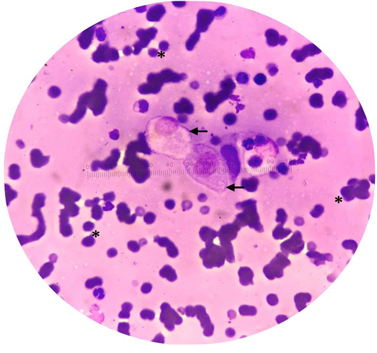
Figure 1
Cytology of impression smear showing erythrocyte (*) and epithelial cells (arrow). (Diff-Quik stain, x40).
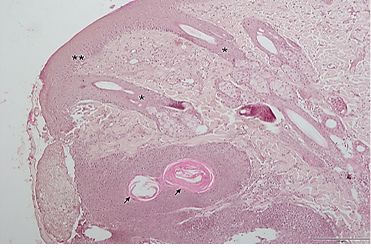
Figure 2
Histopathologic section of the excised tissue: rete accentuation (*), epidermal hyperplasia (**) and keratin pearl (arrow). (H&E, 10x). Scale; 5µm.
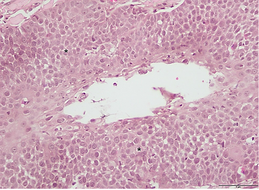
Figure 3
Histologic section of the excised tissue showing epidermal dysplasia (*). (H&E stain, 40x). Scale; 5µm.
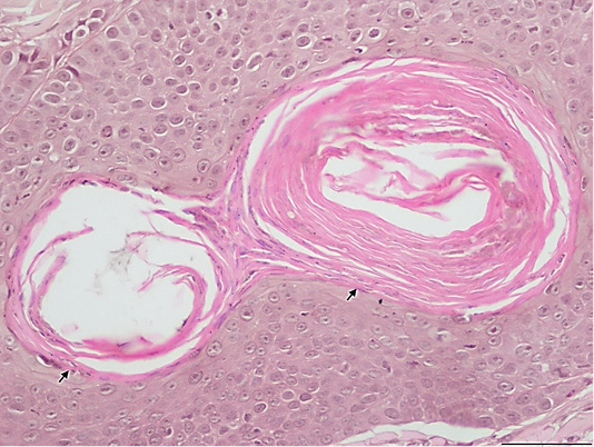
Figure 4
Histologic section of the excised tissue showing keratin pearl (arrow). (H&E, 40x). Scale; 5µm.


