Journal of Animal Health and Production
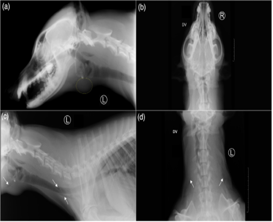
Radiographic views of the skull: left lateral (a) and dorsoventral (b), and the neck: left lateral (c) and dorsoventral (d) of the reported case. (a) There is presence of a rounded soft tissue or fluid opacity structure with distinct margin just ventral to the larynx (marked with yellow dotted circle). (c,d) On both left lateral and dorsoventral radiographs of the neck, multiple small rounded air opacities coalescing to linear gas opacities (white arrows), noticed in bilateral fascial planes of the neck indicating cervical soft tissues emphysema.
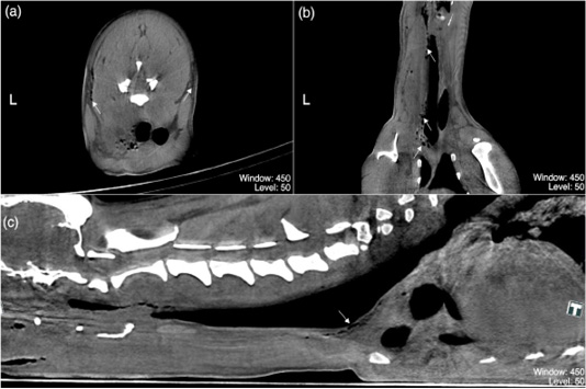
(a–c) are post-contrast multiplanar reformation (MPR) acquisitions of the neck region of the dog in present case study representing (a) transverse plane at the level of C5, (b) dorsal and (c) sagittal planes. Multiple small rounded gas-attenuating densities coalescing to form linear gas–attenuating densities peripheral to the fascial planes, oesophagus, and trachea (white arrows); indicating cervical soft tissues emphysema. Any piece of wood or splinter resembling structures were not identified from these CT acquisitions. (Window: 450; Level: 50)


