Journal of Animal Health and Production
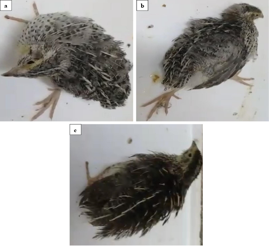
Clinical signs of infected quails with velogenic class II genotype VIb NDV strain (EG/SR/76/CH/1967) at 6 dpi. The infected quails showed torticollis (a), paralysis of the legs (b) and ruffling feather (c).
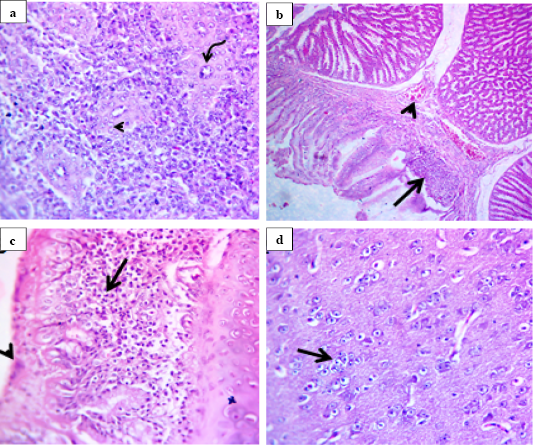
Histological lesions of spleen, proventriculus, trachea and brain of infected quails with velogenic class II genotype VIb NDV strain (EG/SR/76/CH/1967) at 7 dpi (H and E, 400X). Spleen showed pericytoma (arrow head) with hypertrophic epithelium lining the central arteries of white pulp (curved arrow) (a). Proventriculus showed mild congestion of submucosal blood vessels (arrow head) and replacement of necrotic epithelial lining by inflammatory cells (arrow) (b). Trachea showed necrotic respiratory epithelium (arrow head) with subepithelial and submucosal round cell infiltration (arrow) (c). Brain showed replacement of degenerated neuron by glia cells (arrow) (d).
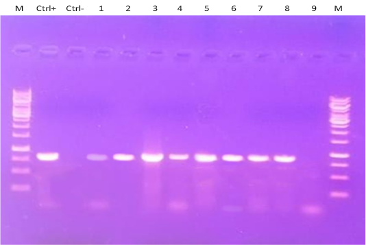
RT-PCR amplification of F gene from clinically infected quails with two NDV strains in the present experiment. Lane M: 1-kbp DNA marker; Ctrl+: control positive; Ctrl: control negative; Lanes 1-6: Amplified products of NDV detected in different tissues of group A that infected with NDV strain (EG/SR/76/CH/1967). Lanes 7-9: Amplified products of NDV detected only in one spleen (lane 7) and one brain (lane 8) tissue samples of group B that infected with NDV strain (NDV/chicken/Egypt/1/2015) at 7 dpi, while lane 9 represented negative NDV infected tissue sample.


