Journal of Animal Health and Production
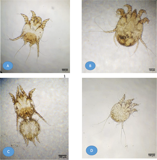
Light microscopy of mite images from buffaloes. (A) Adult Psoroptes natalenesis male; (B) Adult Psoroptes natalenesis female; (C) Adult Psoroptes natalenesis male attached with female tritonymph; (D) Male protonymph Psoroptes natalenesis.
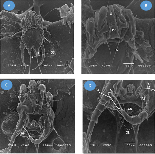
Adult Psoroptes natalenesis male from buffaloes under scanning electron microscope. (A) Dorsal surface; (OS: Opisthsomal setae); (B) Anterior end; Podosoma; (G: Gnathosoma; PP: Propodosomal plate; PS: Propodosomal seta); (C) Ventral surface; (GS: Genital sucker; IV: leg IV (short); III: leg III ended with terminal sucker); (D) Opisthosoma; (MS: Metapodosomal setae; AN: Anus; A: ambulacrum; S: Pulvillus (sucker); OL: Opisthosomal lobes).
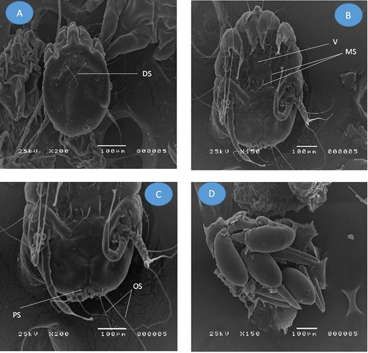
Adult Psoroptes natalenesis female from buffaloes under scanning electron microscope. (A) Dorsal surface; (DS: Dorsal setae); (B) Ventral surface; (V: Vulva; MS: Metapodosomal setae); (C) Opisthosoma; (PS: Perianal seta; OS: Opisthosomal setae); (D) Eggs
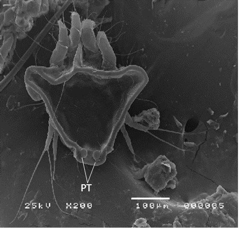
Female protonymph of Psoroptes natalenesis from buffaloes under scanning electron microscope showing dorsoposterior tubercles (PT).
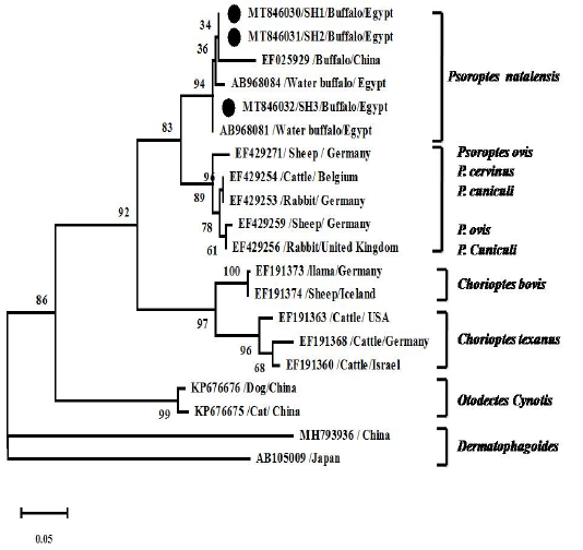
Phylogenetic analyses using Tamura 3-parameter +G as a maximum likelihood models with the lowest BIC scores showing the relationship of the obtained ITS2 gene nt sequences with others closest mite species references. Numbers at nodes show the percentage in 1000 bootstrap replicates.


