Journal of Animal Health and Production
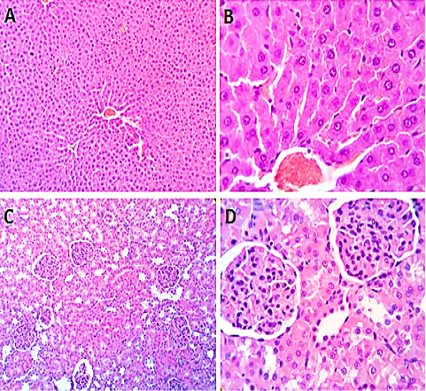
Photomicrograph of control (untreated) group of liver (A and B) showing normal hepatic parenchyma. Kidney (C and D) showing normal renal parenchyma. H&E X 100 (A, C), H&E X 400 (B, D).
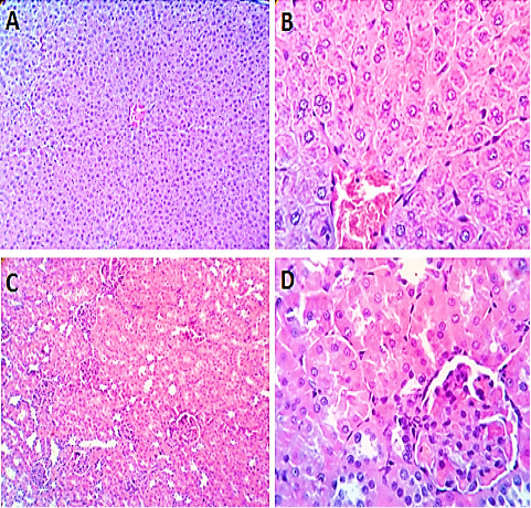
Photomicrograph of vitamin E treated group, of liver (A and B) showing normal hepatic parenchyma with preserved lobular pattern, cord arrangement, vascular tree, sinusoids, kupffur cells and portal area structures. Examined sections from Kidney (C and D) showed normal nephron units with preserved renal papillae, renal pelvis and stroma. H&E X 100 (A, C), H&E X 400 (B, D).
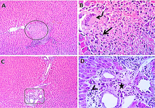
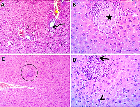
Photomicrograph of Liver of moxifloxacin-treated rats showing multifocal hepatic necrosis of variable sizes (circle), partially replaced by macrophages (arrow) and giant cells (curved arrow). Bile ducts proliferated with characteristic portal round cells aggregation (arrow head) and fibrosis (star) are seen. Some sections showing Centro-lobular degenerative changes in some hepatic lobules and hypertrophied kupffur cells. H&E X 100 (A, C), 400 (B, D).
Photomicrograph of kidney of moxifloxacin-treated rats showing focal sloughing (circle) and hyperplastic changes (black star) in the transitional epithelium of pelvis, focal interstitial and perivascular aggregation of round cells (yellow arrow) and eosinophils (red arrow). Some of the cortical and medullary tubules showed cyst changes. The renal blood vessels showed congestion with mild perivascular edema. A few renal tubules shows degenerative and necrotic changes. H&E X 100(A, C), 400 (B, D).
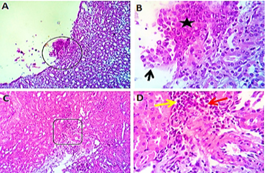
Photomicrograph of moxifloxacin + Vit E treated group, liver showing apparently normal hepatic parenchyma with residual portal biliary proliferation (curved arrow), minute focal hepatic necrotic areas (black star) partially replaced by round cells (open arrow). The hepatic blood vessels showing mild congestion (yellow star) in addition to hypertrophied kupffur cells (arrow head). H&E X 100 (A, C), H&E X 400 (B, D).
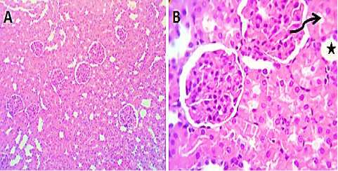
Photomicrograph of kidney of moxifloxacin + Vit E treated group, (A and B) showing apparently normal nephron units with mild degenerative changes in some tubular epithelium (curved arrow) and cyst dilatation of some renal tubules (star). H&E X 100 (A), H&E X 400 (B).


