Journal of Animal Health and Production
Case Report
J. Anim. Health Prod. 9(1): 47-51
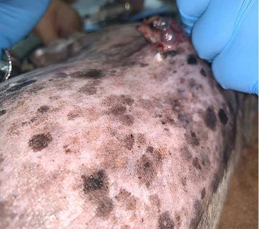
Figure 1
Generalized brown-greyish and blackish macules of varying sizes with irregular borders.
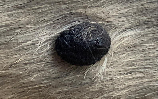
Figure 2
Focal and soft black nodules with a smooth surface of 1 cm in size observed at the right lateral trunk region.
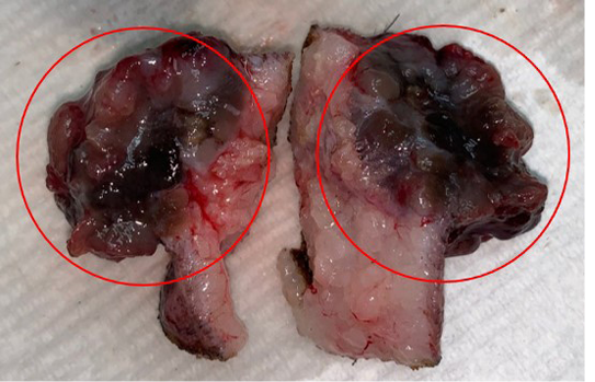
Figure 3
Brownish-black mass with cauliflower-like appearance (circles).
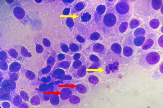
Figure 4
Pleomorphism characterized by a mixture of spindle and round cells and presence of multiple nucleoli (red arrows), mitotic figures (yellow arrows) and intracytoplasmic melanin granules (H and E, 1000X).
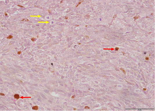
Figure 5
Pleomorphism with spindle and round cells, anisokaryosis and mitotic figures (yellow arrows) as well as intracytoplasmic melanin pigments observed within the dermis (red arrows) (H and E, 400X).
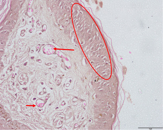
Figure 6
Pagetoid melanocytosis (circle) and evidences of angiogenesis (arrows) (H and E, 400X).


