Journal of Animal Health and Production
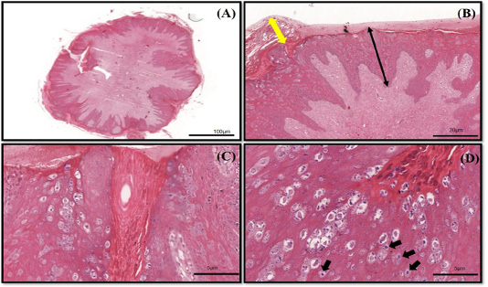
The histopathology findings of bovine papilloma tissues in this case. (A) Low magnification showing a well-developed finger-like projecting papillae rising from the surface of the epidermis to the subcutaneous layer. (B) Hyperkeratosis of the epithelial layer (double yellow arrow) with acanthosis formation (double black arrow). (C) Epidermal proliferation with elongated rete pegs and neoplastic fibroblast. (D) Multiple koilocytosis formation on the dermis layer (black arrows). H&E staining 10X and 40X.
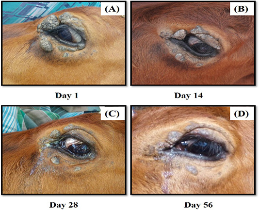
The progression of treatment after administration of autogenous BPV vaccine. There was marked regression of the tumour growth from (A) Day 1, (B) Day 14, (C) Day 28 and (D) Day 56.
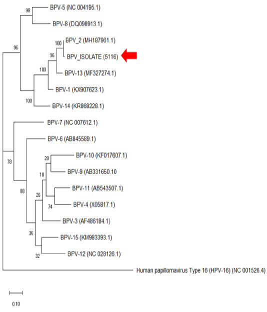
The phylogenetic analysis of the genome sequence of bovine papillomaviruses using the maximum-likelihood method by 1,000 bootstrap replications. A positive sample in this study is indicated by an arrow.


