Journal of Animal Health and Production
Case Report
J. Anim. Health Prod. 8(3): 145-149
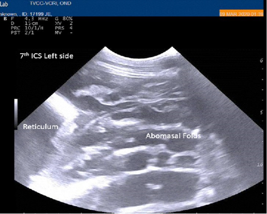
Figure 1
Percutaneous ultrasonography of left lateral thorax at 7th inter coastal space showed inflamed abomasal leaves with emptiness.
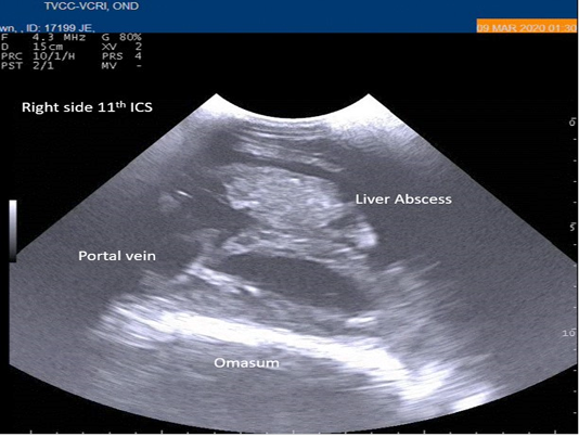
Figure 2
Percutaneous ultrasonography of right-side abdomen at 11th inter coastal space showed liver hyperechoic foci with anechoic cavity.
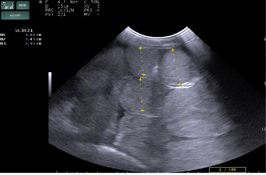
Figure 3
Percutaneous ultrasonography of right side mid abdomen showed dilated (>4.5cm) intestinal loops with mild peritonitis (Arrow).
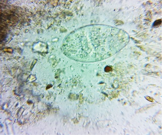
Figure 4
Faeces examination by sedimentation method showed Amphistome eggs (x 400) in three animal.
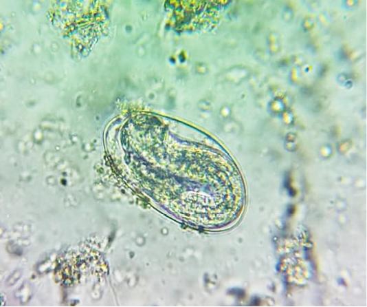
Figure 5
Faeces examination by sedimentation method showed Strongyloides eggs (x 400) in one animal.


