Journal of Animal Health and Production
Case Report

Dystocia Handling by Cesarean Section in Beetal Goat in Pakistan: A Surgical Approach
Nasir Iqbal1*, Sadaf Aslam1, Naveed Hussain1, Zubair Luqman2, Hamza Jawad2
1Department of Veterinary Surgery & Pet Sciences, University of Veterinary and Animal Sciences, Lahore, Punjab, Pakistan; 2Faculty of Veterinary and Animal Sciences, The Islamia University of Bahawalpur, Bahawalpur, Punjab, Pakistan.
Abstract | Dystocia in Beetal goat breed is common due to keeping of mixed herd of different breeds at the same place. A vaginal examination should not practice unless required, as it may result in drastic effects over kid and goat as well. A non-descriptive pregnant Beetal goat was presented with the signs of left fore-limb protruding out since 2 days from the vulva. On ultrasonographic examination, it was concluded that the dystocia was due to fetal maternal disproportion. The caesarean section was performed under complete aseptic conditions to avoid any post-operative infection. The left flank approach was used under inverted L block on left Para lumber fossa by using 2% Lignocaine. Alive fetus with an intact umbilical cord was drawn out from the uterus of the goat. Suturing was performed as per standard surgical methods and post-operative care was done including antiseptic dressing of wound and pain management for five days. It is suggested that separate breed herd management should be a practice to avoid such life-threatening conditions.
Keywords | Beetal goat, Dystocia, Caesarean, Aseptic, Suture
Received | March 29, 2020; Accepted | May 27, 2020; Published | July 26, 2020
*Correspondence | Nasir Iqbal, Department of Veterinary Surgery & Pet Sciences, University of Veterinary and Animal Sciences, Lahore, Punjab, Pakistant; Email: nasir.iqbal@uvas.edu.pk
Citation | Iqbal N, Aslam S, Hussain N, Luqman Z, Jawad H (2020). Dystocia handling by cesarean section in beetal goat in pakistan: a surgical approach. J. Anim. Health Prod. 8(3): 134-137.
DOI | http://dx.doi.org/10.17582/journal.jahp/2020/8.3.134.137
ISSN | 2308-2801
Copyright © 2020 Iqbal et al. This is an open access article distributed under the Creative Commons Attribution License, which permits unrestricted use, distribution, and reproduction in any medium, provided the original work is properly cited.
Introduction
Dystocia is defined as the failure of transmission of stage one to stage two labor or when thirty minutes spend to start of stage two of labor or parturition (Ahmed et al., 2017; Kumar et al., 2016). Stage one of parturition (labor) is defined as the preparation for fetal expulsion and includes decreased appetite, preparing a birthing area, isolation from the herd, restlessness, and leading up to the early uterine contractions. (Majeed and Taha, 1989). Fetal expulsion or delivery of neonate is stage two of parturition. In small ruminant’s dystocia occur because of fetal-maternal disproportion, multiple fetuses within the pelvic canal, fetal malpositioning, incomplete dilation of the cervix, and uterine inertia (Brounts and Serbina, 2004; Dalal et al., 2017; Reddy et al., 2016). Dystocia because of fetal-maternal disproportion occurs when a heavy male breed mated with a small female breed (Hafez et al, 2013; Tripathi et al., 2016). In this condition, birth of a fetus does not occur through the birth canal because of the giant size of a fetus or newborn and small size of the birth canal (Tripathi et al., 2016) . Caesarean is the only handling of fetal-maternal dystocia followed by systemic administration of antibiotics and non-steroidal anti-inflammatory drugs. Single layer cesarean section closure must be done in two layers in spite of single layer closure as it confirms zero leakage after checking uterine leakage. Caesarean or c-section is less required in sheep and goat than cattle, because of the lesser frequency of fetal-maternal disproportion as basis of dystocia i.e. only 1 out of 5 compared with 1 out of 2 in cattle (Al-Timimi, 1997; Kachiwal, 2000; Bhattacharyya et al, 2015).
Case History and Clinical Management
Beetal goat, aged about 2.5 years and 23 kg bodyweight with the history of partial one left fore-leg out from vulva since 48 hours was presented at our clinic in Rahim Yar Khan, Punjab, Pakistan. The animal was suffering from this condition for over past three hours. The owner and veterinary assistant tried to push the fetus manually, but unfortunately, they did not pull it out from the birth canal (Figure 1).
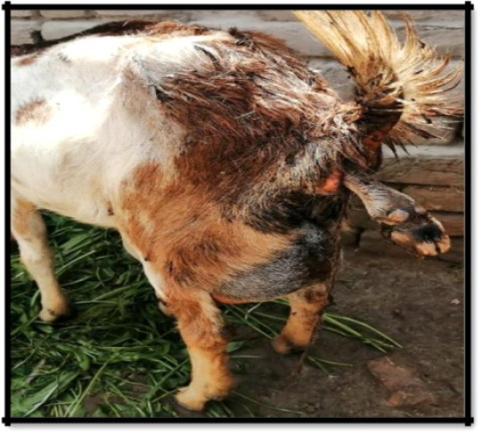
Figure 1: Partial one left fore-limb of fetus protruding out from the vulva of beetal dam (Red Arrow)
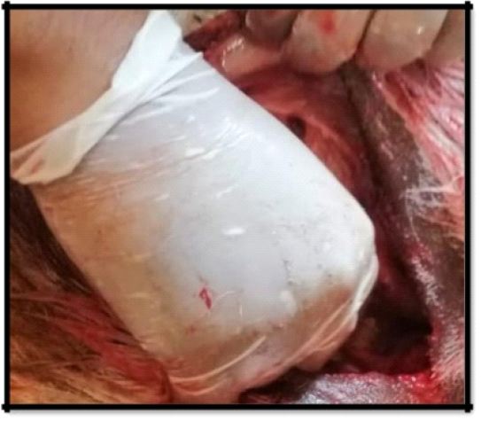
The owner told that they kept a different breed of animals in the same herd and Rajanpuri buck (male goat) sexually mated with Beetal doe; female goat (Phillip et al., 1985). On clinical examination, body temperature was 102⁰C with normal respiration and tachycardia and there was no injury and hemorrhage over limb. Animal dehydration status is normal, checked by skin fold test or skin turgor test, but the animal was restless. The fetus was alive as per checked or confirmed by their reflexes. The presentation, posture, and position of the fetus were normal but did not come out from the pelvic canal due to their oversize (Majeed and Taha, 1993). Ultrasound examination confirmed large size fetus so it was decided to perform Cesarean. After proper restraining, the Beetal goat was placed on the right lateral recumbency exposing the left flank (Gupta and Chhavi, 2020; Robert and Stephan, 1986; Al-Kass and Basheer, 2005). Clipped the hairs on left para-lumbar fossa by using trimmer and injected 0.05 mg/kg xylazine intramuscularly for mild sedation (Kachiwal, 2000). After that, the surgical site was scrubbed with pyodine (10% povidone-iodine) as an antiseptic and used lidocaine (2% lignocaine) for inverted L-block on left para-lumbar fossa as a local anesthetic agent (Majeed and Taha, 1994). About 20 cm long vertical incision was placed on left para-lumbar fossa (skin), approximately10-12cm below the transverse process with the help of a surgical blade. The approach was continued by means of a combination of blunt and sharp dissection over subcutaneous tissues and abdominal muscle layer to approach the abdominal cavity (Majeed and Taha, 1989). Uterine horn of gravid uterus was grabbed and exteriorized gently to avoid perforation and placed holding suture on the uterine horn for holding it outside the abdominal cavity (Figure 2).
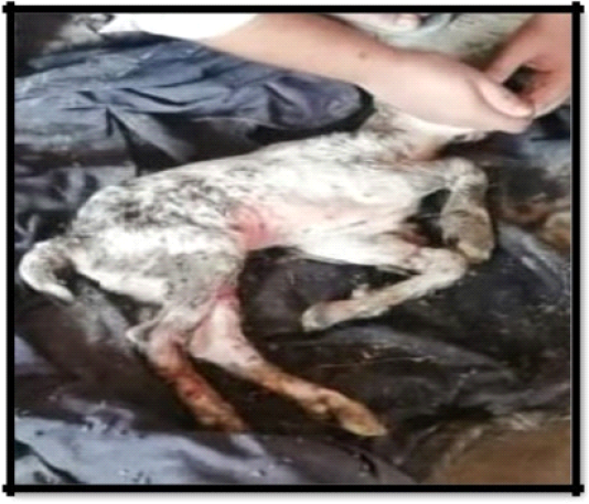
Hysterotomy (incision in the uterus) was performed on the greater curvature of gravid horn and removed fetus from the uterine horn by grasping posterior limb of the fetus with intact umbilical cord (Hussain and Zaid, 2010). The fetus was detached from the umbilical cord and nostrils were cleared from fluid to facilitate proper respiration (Figure 3).
Single layer closure has been done by using Chromic Catgut number 2 with Cushing suture pattern and perforation has been checked by uterus leaking technique and surprisingly due to 1.5mm suture gap there was no leakage found from the uterus and animal has been recovered quite efficiently. Uterus was washed to remove blood clots and foreign particles with normal saline (Majeed and Taha, 1992). Penbiotic (Nawan Laboratories) having Procaine Penicillin, Benzyl Penicillin and Streptomycin Sulphate of about 5 grams was poured in uterus and in abdominal cavity to prevent secondary bacterial infection. Greater curvature of uterine horn was enclosed by using absorbable suture material chromic cat gut number 2 single layer by Cushing suture pattern with 1.5mm sutures gap. Simple interrupted suture on the muscles layer and simple continuous suture on a subcutaneous layer with chromic catgut number 2. Simple interrupted suture pattern was applied on the skin with silk number 2 suture material (Figure 4).
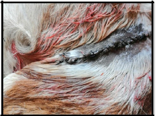
Figure 4: Enclosed skin by using Silk
Antiseptic dressing was applied on sutures for about 7 days (Brounts and Serbina, 2004). The animal was returned to its normal routine within 10 hours after surgery. Loxin (Selmore Pharma) having a nonsteroidal anti-inflammatory drug flunixin meglumine was administered intramuscularly (IM) at the dose rate of 2 mg/kg for a period of about five days to avoid swelling and get relief from pain. Nawacin 50 (Nawan Laboratories) having antibiotic tetracycline at the dose rate of about 20 mg/kg was given to prevent from secondary infection for five days intramuscularly (Hafez et al., 2013). There was no need of statistical analysis as it has been done in a single animal.
Results and Discussion
After 3 to 4 hours post operatively the animal was completely active and there were no signs of internal or external bleeding. Post operative care and anti septic dressing was done for a period of five days to avoid any secondary bacterial infection (Phillip et al., 1985). Sutures were opened on day 10 postoperatively by using suture cutting needle resulting into proper and clear adhesion was there after suture removal (Majeed and Taha, 1994; Ismail and Zuhair Bani, 2017).
Dystocia due to fetal-maternal disproportion in small ruminants is a common clinical case due to different sized mix breed herding at a same place that has been successfully managed by cesarean followed by using Broad spectrum antibiotic i.e. from group Tetracycline and non-steroidal anti-inflammatory drug i.e. Flunixine Meglumine therapy and the same was depicted by (Ahmed et al., 2017; Majeed and Taha, 1994). If cesarean did not occur in time, it will be fatal for damn and fetus life as it may results into death of fetus and ultimately mummification and maceration steep toward the low body condition score of animal resulting into death of goat by systemic infection same as told by (Ahmed et al., 2017; Ismail and Zuhair Bani, 2017; Gupta and Chhavi, 2020; Kachiwal, 2000). This type of dystocia mainly occurred due to the sexual matting of large and small breeds and same has been happened in this case. For the prevention of this sort of dystocia, it is necessary to keep the same type of breeds in a herd instead of mixed breeds herd at a same place.
Conclusion
Single layer closure of uterus with the suturing gap of about 1.5mm can also result into normal healing for cesarean section. Dystocia due to fetal maternal disproportion is common in different size mix breed herd at a same place that can be overcome by separate herding of same size breed at an individual place.
conflict of interest
There is no conflict of interest.
authors contribution
Nasir Iqbal, Naveed Hussain, Sadaf Aslam: Experimental Trial.
Zubair Luqman, Hamza Jawad: Formatting, Setting and Revision.
References


