Journal of Animal Health and Production
Case Report

Mucohaemorrhagic Enteritis Caused by Mixed Parasitic Infection in a Large White Yorkshire Pig
Saravanan M1*, Ramkumar PK1, Rani N3, Kannan K1, Selvaraj P4, Venkatesan M2, Yogespriya S2, Jayalakshmi K2, Senthilkumar S1
1Veterinary Clinical Complex; 2Department of Veterinary Medicine, VCRI Orathanadu; 3Department of Veterinary Parasitology, VCRI Namakkal. 4Department of Veterinary Medicine, Madras Veterinary College, Chennai -7.
Abstract | Mixed parasitic infection is a challenging task in terms of diagnosis and therapy and unidentified cases may lead to life threatening situation in humans and animals causing zoonosis. Here author report a case of a four month old male large white Yorkshire pig with the history of inappetence and chronic diarrhoea which is hemorrhagic in nature at its lateral stages of illness. Clinical, faecal examination and Egg per gram (EPG) studies were done to identify the presence of parasites. Large numbers of motile Balantidium coli, motile Ascaris suum larva and Ascaris suum eggs were seen under microscopic examination. EPG studies showed high counts for Trichuris suis, and Ascaris suum. Hematology revealed anemia and leukocytosis. Medical management consisting of fluid therapy, anti parasites and supportive therapy were given and pig was recovered uneventfully two weeks of treatment.
Keywords | Ascaris suum, Balantidium coli, Swine, Trichuris suis, Zoonosis
Received | March 29, 2020; Accepted | May 27, 2020; Published | July 26, 2020
*Correspondence | Saravanan. M, Veterinary College and Research Institute, Orathanadu, Thanjavur - 614 625; Email: sara82vet@yahoo.com
Citation | Saravanan M, Ramkumar PK, Rani N, Kannan K, Selvaraj P, Venkatesan M, Yogeshpriya S, Jayalakshmi K, Senthilkumar S (2020). Mucohaemorrhagic enteritis caused by mixed parasitic infection in a large white yorkshire pig. J. Anim. Health Prod. 8(3): 130-133.
DOI | http://dx.doi.org/10.17582/journal.jahp/2020/8.3.130.133
ISSN | 2308-2801
Copyright © 2020 Saravanan et al. This is an open access article distributed under the Creative Commons Attribution License, which permits unrestricted use, distribution, and reproduction in any medium, provided the original work is properly cited.
Introduction
In developing countries like India, majority of pigs are raised under free-ranging system where they feed with raw garbage and kitchen waste, therefore more prone to parasitic infections (Tiwari et al., 2009; Weka and Ikeh, 2009). Around 90% of the pig population in India are in rural areas, where swine domestication is mainly concentrated to low income group families having poor hygiene standards (Laha et al., 2014). Helminthic infection in pigs were results reduce feed conversion and decreases carcass yield, which leads huge economic losses (Boes, 2000; Harper, 2004). The prevalence of gastrointestinal parasites in pigs are widely reported from all over the world by many workers, viz., Ascaris suum, Trichuris suis (Manuel et al., 1989; Dutta et al., 2005). In addition to helminths, the pigs also harbour many intestinal protozoan, viz., Balantidium coli in developing countries (Patra et al., 2019). Here, such a parasitic infection was reported as Mucohaemarrhagic enteritis due to concomitant infection of Trichuris suis, Ascaris suum and Balantidium coli in Large White
Yorkshire Pig.
Case History and Observations
A four months old male Large White Yorkshire pig from a flock of 23 piglets of semi intensive rearing with no proper deworming was referred to the Large Animal Medicine Referral Clinic, VCRI, Orathanadu with the history of diarrhoea for past ten days later hemorrhagic (Figure 1) in nature for consequent three days and inappetant. On clinical examination all the vital parameters were normal. Faecal examination revealed large numbers of motile Balantidium coli (Figure 2), motile Ascaris suum larva (Figure 3) and Ascaris suum eggs (Figure 4) and Trichuris suis eggs (Figure 5). Adult worms of Ascaris suum (Figure 6) were also expulsed from the diarrhoeic faeces. Faecal sample subjected to quantification of egg count revealed 1100 EPG of Trichuris suis eggs and 900 EPG of Ascaris suum eggs. Hemato-biochemical changes were analyzed and depicted in Table 1. Anemia and leukocytosis were observed.
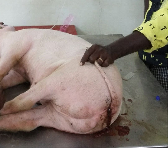
Figure 1: Muco haemorrhagic diarrhoea in a pig
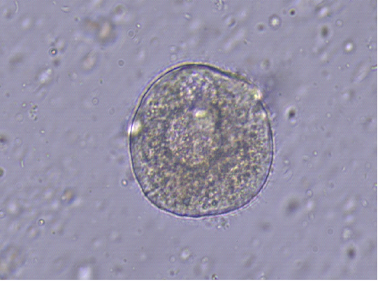
Figure 2: Balantidium coli with cilia (40x)
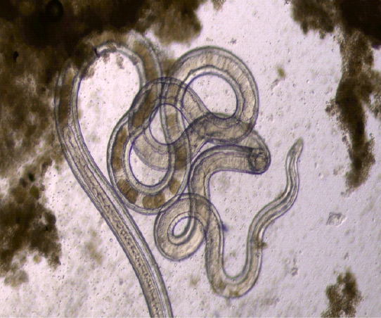
Figure 3: Ascaris suum motile larva (4x)

Figure 4: Ascaris suum egg (40x)
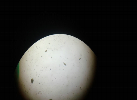
Figure 5: Trichuris suis eggs (10x)
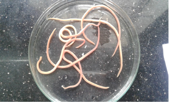
Figure 6: Adult worms of Ascaris suum
Treatment & Management
During the first day of treatment, the fluid and electrolyte loss was corrected with intravenous Inj. Ringers lactate @ 10ml/kg bwt followed by Inj. Metronidazole @ 20 mg/kg bwt IV, Inj. Chlorpheniramine maleate @ 0.5 mg/kg bwt IM and Inj. B complex @ 1ml were administered. To counteract the parasitic load, an oral anthelmintic Susp. Levamisole and Rafoxanide @ 1ml/ 4kg bwt were administered. Oral amino acid and haematinics were given as supportive therapy to correct anemia. The owner was advised to keep the affected pig on isolation. Next day, the animal voided numerous dead Ascarid larva in diarrheic faeces. After four days of treatment, EPG count becomes nil and pig gradually improved in terms of activity and appetite. The other pigs are treated with appropriate anthelmintic and measures were given for the proper disposal of faeces to prevent further occurrence in the pig shed area.
Considering the zoonotic potential of the present disease, the farm managers must take proper advice from physician working in human hospital regarding the choice of anthelmintic for human beings.
Table 1: Hemato-biochemical alteration in pig due to concomitant infection of Trichuris suis, Ascaris suum and Balantidium coli.
| Parameters | Value |
Reference range (Constable et al., 2017) |
| Hemoglobin (g/dl) | 6.5 | 10-16 |
| PCV (%) | 18 | 32-50 |
|
RBC (x 106/µl) |
2.01 | 5-8 |
|
WBC (x 103/µl) |
28.01 | 11-22 |
| Total Protein (g/dl) | 5.2 | 4.5- 7.3 |
| Albumin (g/dl) | 2.35 | 1.9 - 4.0 |
| Urea (mg/dl) | 29 | 10 - 30 |
| Creatinine (mg/dl) | 1.22 | 1 - 2.7 |
| AST/SGOT (U/l) | 43 | 32- 84 |
PCV: Packed cell volume; RBC: Red blood cell; WBC: White blood cell; AST: Aspartate aminotransferase; SGOT: Serum glutamic-oxaloacetic transaminase
Discussion
The clinical signs associated with Ascaris suum, the largest roundworm of the pig includes dyspnoea, increased depth of respiration, coughing, diarrhoea and jaundice and the pathology of the disease is mainly due to migrating larvae in the visceral organs (Roepstorff and Nansen, 1998). Trichuris suis (whip worm) causes mucoid diarrhoea, anemia, dehydration, poor growth and emaciation in pigs. Being a normal commensal in pig, Balantidium coli under opportunistic infection will cause dysentery, general malaise and depression. The differential diagnosis of mucohaemorrhagic diarrhoea in swine is Swine dysentery. Anemia and leukocytosis of this present cases due to hemorrhagic diarrhea and secondary bacterial infection. The main clinical feature reported in the present case is mucohaemorrhagic diarrhoea which might be due to inflammation of the small and large intestine especially caecum and colon as reported earlier by other researcher and this is associated with Ascaris suum, Trichuris suis and Balantidium coli (Roepstorff andNansen, 1998). As per the available literature, Ascaris spp. and Trichuris spp. are the most prevalent helminthic infections in humans and pigs worldwide (Holland and Boes, 2002) whereas A.suum is commonly recorded in Punjab (Kaur et al., 2017). The worms that infect pigs and humans are closely related and difficult to distinguish morphologically owing to a lack of discrete characters. Pigs harboring Ascaris suum and Balantidium coli can act as potential source of human health hazards (Chawhan et al., 2014). In some region, pig manure is used as fertilizer for the cultivation of vegetables meant for human consumption leading to development of cross-infections which is considered as potential risk factor (Nejsum et al., 2012). Hence, proper cocking of the pork before eating may helps to prevent helminthic infestation via parasitic eggs and cysts (Gagman et al., 2014). This disease could be the sentinel for future mortality. As Ascaris suum and Balantidium coli are highly zoonotic, animal handlers were advised to take proper anthelmintic as per physician recommendation.
The mixed parasitic infections are quite challenging and detrimental effects on pigs and human health. Hence, proper disposal of the faeces and sanitary measures should be taken for complete eradication of the parasitism in the soil / environment.
Acknowledgments
The Authors are grateful to The Director of Clinics TANUVAS and The Dean VCRI, Orathanadu to provide all the facilities for carrying out this study.
Conflict of interest
The presented work is purely a clinical case and there was no conflict of interest.
authors contribution
Saravanan M: Case investigation, literature collection, Manuscript preparation and Manuscript corresponding.
Ramkumar PK: Case treatment and sample collection.
Rani N: Parasitology sample processing and interpretation.
Kannan K: Result compilation.
Selvaraj P: Treatment suggestion.
Venkatesan M: Hematology analysis.
Yogespriya S: Biochemical analysis.
Jayalakshmi K: Parasitology sample processing interpretation.
Senthilkumar S: Manuscript proof reading.
References


