Journal of Animal Health and Production
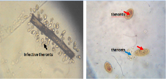
Infective theronts (black arrow) attached to a substrate and tomonts actively releasing theronts. Lugol iodine stain, mag. X40.
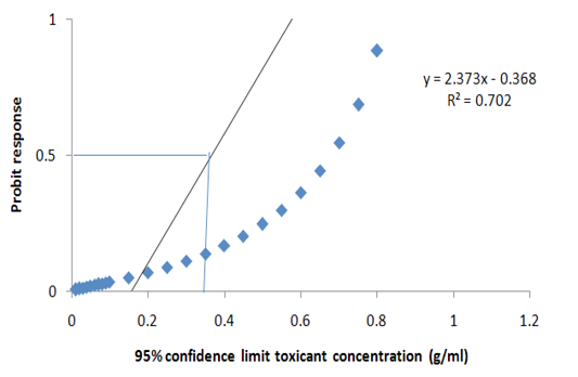
Probit transformed responses for minimum inhibitory concentration in the antiprotozoal activity of M. oleifera on the key life stage (theronts) of Ichthyophthirius multifiliis.
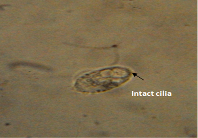
Normal morphology of theronts showing intact cilia (black arrow) with fusiform shape. Neutral red stain. mag. X40.
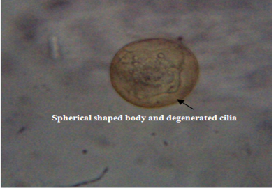
Deformed theronts after exposure to aqueous leaves extract of Moringa oleifera showed, distorted and degenerated cilia and spherical shaped body (black arrow). Neutral red stain. mag. X40
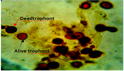
Gill of fish showing dead (red arrow) and live (black arrow) trophonts with intact nucleus under lugol iodine stain during treatment with aqueous leaves extract of Moringa oleifera.
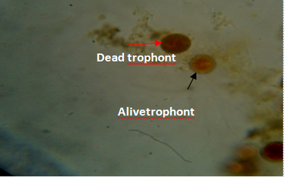
Body smear of fish showing dead (red arrow) and alive trophonts (white arrow) with intact nucleus under lugol iodine stain during treatment with aqueous leaves extract Moringa oleifera. Mag. 100X.


