Journal of Animal Health and Production
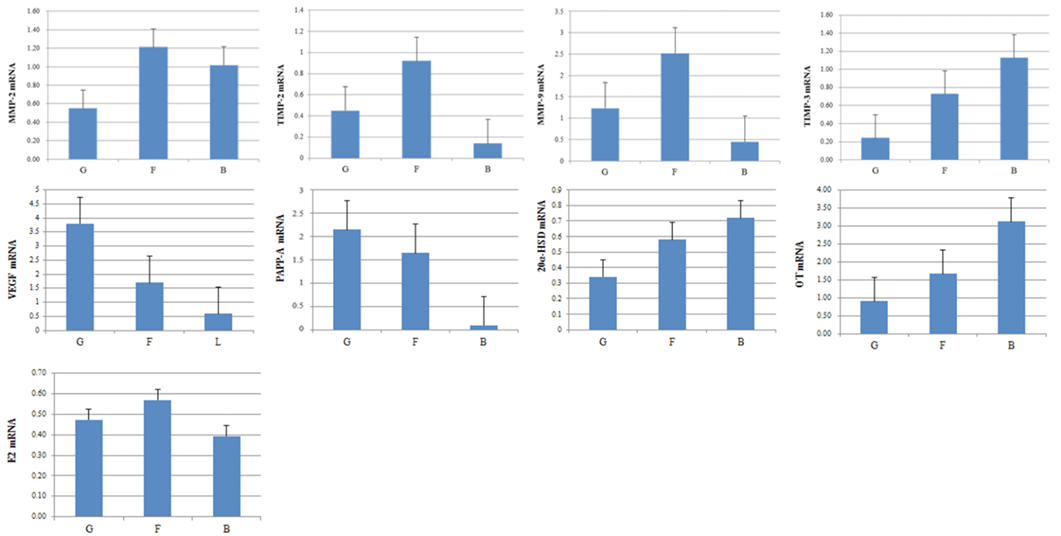
Expression of pregnancy-associated plasma protein A, VEGF, 20-Hydroxysteroid dehydrogenase, OT, E2, MMP-2, MMP-9, TIMP-2 and TIMP-3 mRNA in EECs. Expression of embryo quality-associated mRNAs in the EECs. Samples from good- (G), fair- (F), and low-quality group (B).
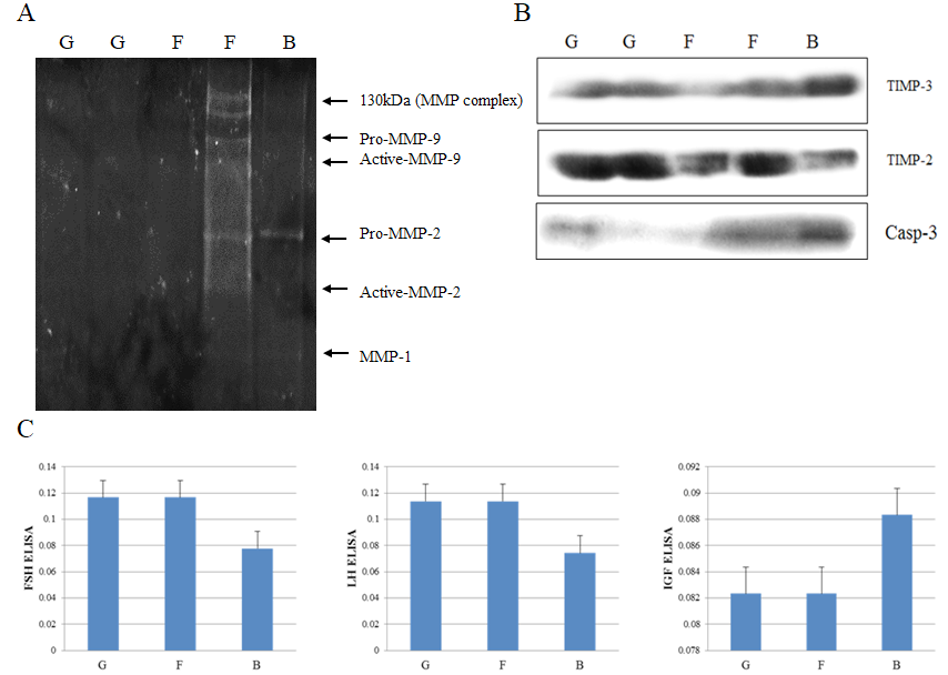
Protein expression analysis of MMP-2, MMP-9, TIMP-2, TIMP-3 and Casp-3. Gelatin zymography (A) of embryo quality in uterine milk. Western blot (B) of embryo quality in EECs. ELISA-based analysis of FSH, LH, and IGF concentration in the serum of good-, fair- and low-quality groups (C). Samples of good- (G); fair- (F) and low-quality group (B).
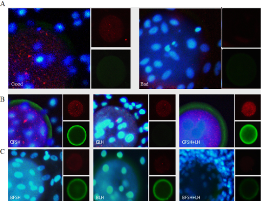
Immunofluorescence analysis of embryo quality-related proteins in embryos and EECs incubated for 24 h. Expression of PCNA and pregnancy-associated plasma protein A (PAPP-A) in the embryo and EECs of the good (Good) and low-quality (Bad) groups (A). The large panel shows the merged picture. Grading of EECs: Good- (B) and low-quality (C) groups. GFSH: FSH-treated embryos and EECs; GLH: LH-treated embryos and EECs; GFSH+LH: FSH + LH-treated embryos and EECs. Embryos were developed in the absence or presence of hormones. The large panel shows the merged picture. The small panel shows the protein detected (red: PCNA), (green: PAPP-A).
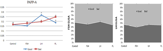
ELISA-based analysis of pregnancy-associated plasma protein A, FSH and LH in the embryo and EEC culture conditioned media. Samples of good- (blue) and low-quality groups (red). (con: control group; FSH: follicle stimulating hormone; LH: luteinizing hormone; FL: follicle stimulating hormone + luteinizing hormone).


