Journal of Animal Health and Production
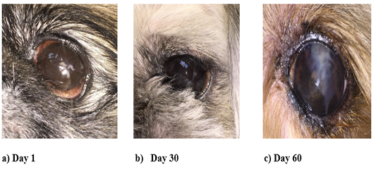
Visual changes in the anterior surface of the eye in animals of the control group with moderate KCS
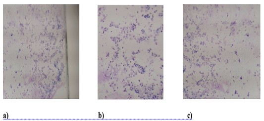
Cytology of the front surface of the eye in animals of the control group with moderate KCS.
a) (Day 1), b) (Day 30) and c) Day 60, have over 50 cells each (epithelial cells and segmented cells). The background is slimy.
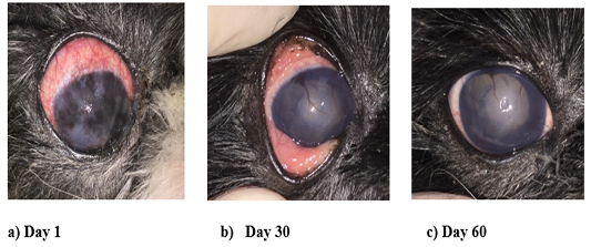
Visual changes in the anterior surface of the eye in animals of the experimental group with moderate KCS
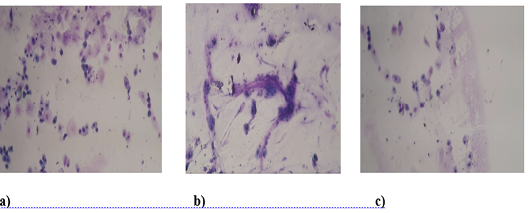
Cytology of the anterior surface of the eye in animals of the experimental group with moderate KCS.
a) Day 1. High cellularity. It is mainly represented by epithelial cells and segmented cells. b) Day 30. Mucous background, cellularity moderate. Epithelial cells and segmented cells are presented. c) Day 60. Cellularity is not high. Mostly epithelial cells.


