Journal of Animal Health and Production
Case Report
J. Anim. Health Prod. 7(4): 166-170
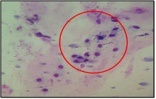
Figure 1
Cytology of the submandibular mass showing a few clusters of round to spindle shape cells with coarse chromatin embedded in pink cellular matrix.
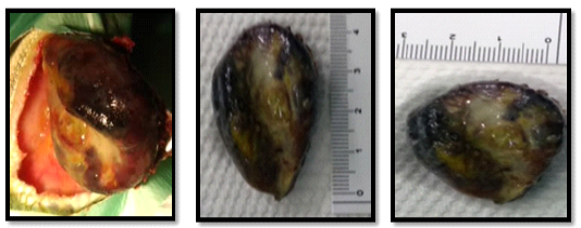
Figure 2
Dimensions of the resected mass (4 cm x 3 cm) with firm and smooth surface.
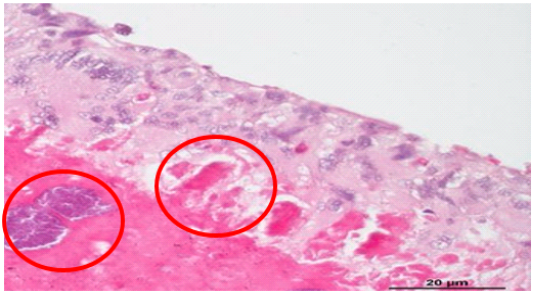
Figure 3
Histopathologic findings showing bony mass with minimal narrow space (x40)
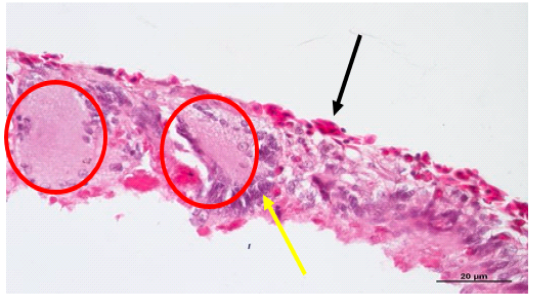
Figure 4
Histopathological findings showing lamellar trabecular-likes structures with outer basophilic and flattened cells.
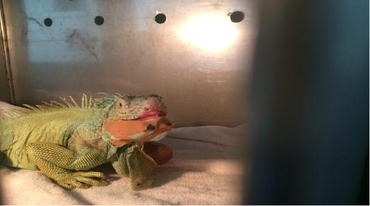
Figure 5
Iguana after removal of the submandibular mass


