Journal of Animal Health and Production
Short Communication

Incidences and Veterinary Clinical Management of Postpartum Diseases Among Domestic Cows and Does in Klang Valley, Malaysia
Faez Firdaus Abdullah Jesse1,2,3, Innocent Damudu Peter1,4, Eric Lim Teik Chung2,5, Nuriza Tukiran1, Asinamai Athliamai Bitrus6, Idris Umar Hambali4, Bura Thlama Paul1, Nur Azhar Amira1, Mohd Azmi Mohd Lila7
1Department of Veterinary Clinical Studies, Faculty of Veterinary Medicine, Universiti Putra Malaysia, 43400 UPM Serdang, Selangor, Malaysia; 2Institute of Tropical Agriculture and Food Security, Universiti Putra Malaysia, 43400 UPM Serdang, Selangor, Malaysia; 3University Community Transformation Centre (UCTC), Universiti Putra Malaysia, 43400 UPM Serdang, Selangor, Malaysia; 4Faculty of Veterinary Medicine, University of Maiduguri, P.M.B 1069, Maiduguri, Nigeria; 5Department of Animal Science, Faculty of Agriculture, Universiti Putra Malaysia, 43400 UPM Serdang, Selangor, Malaysia; 6Research Unit, Microbial Food Safety and Antimicrobial resistance, Department of Veterinary Public Health, Faculty of Veterinary Science, Chulalongkorn University, 10330 Pathumwan Bangkok, Thailand;7Department of Veterinary Microbiology and Pathology, Faculty of Veterinary Medicine, Universiti Putra Malaysia, 43400 UPM Serdang, Selangor, Malaysia.
Abstract | The postpartum period is a time that is characterised by an increased risk of developing parturition related diseases and disorders by the dam. These may affect production in affected animals. The objective of this study was to determine the incidences and methods of clinical management of postpartum diseases among farm animals within the Klang Valley in Selangor, Malaysia. Primary data were obtained from a four-year (January 2013 to December 2017) farm record from sixteen (16) cattle and goats mixed farms. Postpartum diseases and disorders made up 3.4% (53/1550) of total diseases and disorders recorded for cows and does. Out of the 53 cases of postpartum diseases and disorders recorded, the followings were observed; metritis (24.5%), vaginal prolapse (22.6%), pyometra (20.8%), retained placenta (15.1%), uterine prolapse (9.4%), endometritis (5.7%) and uterine tears (1.9%). Most of the recorded postpartum cases were seen in cattle (83.0%) than in goat (17.0%) populations. More postpartum diseases and disorders were seen in younger cows (56.8%) as compared to older cows (43.2%). Similarly, more cases were seen in older does (66.6%) as compared to younger does (33.4%). Furthermore, a higher frequency of postpartum diseases was observed in animals during their first parturition (77.4%) than in animals during their second (17.0%) or third parturition (5.6%). Flunixin meglumine and oxytetracyclines administered via the uterus were frequently used in clinical management of postpartum diseases. However, a detailed and strict follow up of the cases were lacking (52.8%). This study shows that postpartum diseases are quite common in primipara ruminants in the Klang Valley. Although conventional methods of managing postpartum cases in the Klang valley yielded satisfactory outcomes, there is need for compliance on detailed follow up therapy to avoid cases of treatment failures in affected animals.
Keywords | Clinical management, Klang valley, Postpartum diseases, Prevalence, Ruminants.
Received | June 20, 2019; Accepted | August 02, 2019; Published | September 15, 2019
*Correspondence | Faez Firdaus Abdullah Jesse, Department of Veterinary Clinical Studies, Faculty of Veterinary Medicine, Universiti Putra Malaysia, 43400 UPM Serdang, Selangor, Malaysia; Email: jesse@upm.edu.my
Citation | Jesse FFA, Peter ID, Chung ELT, Tukiran N, Bitrus AA, Hambali IU, Paul BT, Amira NA, Lila MAM (2019). Incidences and veterinary clinical management of postpartum diseases among domestic cows and does in klang valley, malaysia. J. Anim. Health Prod. 7(3): 113-118.
DOI | http://dx.doi.org/10.17582/journal.jahp/2019/7.3.113.118
ISSN | 2308-2801
Copyright © 2019 Jesse et al. This is an open access article distributed under the Creative Commons Attribution License, which permits unrestricted use, distribution, and reproduction in any medium, provided the original work is properly cited.
Introduction
The resumption of normal oestrus cycle after parturition is vital for the restoration of breeding potentials in animals. Postpartum diseases and disorders have been reported to cause significant economic losses to farmers by extending the resumption to normal oestrus cyclicity in animals and a longer calving intervals (Kawashima et al., 2006; Walsh et al., 2007; Crowe, 2008; Vergara et al., 2014; Lucy, 2017; Canadas et al., 2019). An increased duration of oestrous cycle and an increased calving interval due to postpartum related diseases are common reproductive problems in livestock especially in smallholder farms (Crowe, 2008; Lucy, 2017). This is of economic importance as affected animals are often culled thereby causing losses to the farmer (Vergara et al., 2014). Prompt diagnosis followed by an effective therapy to postpartum diseases in livestock minimizes and therefore controls the impact of such diseases (Abdullah et al., 2014a). Retrospective studies provide valuable information about the occurrence and pattern of diseases in a population. It is also useful in the assessment of the outcomes of how interventions played out. The objective of this study is to determine the incidence of postpartum diseases and disorders in cow and doe populations in semi intensively managed ruminant farms within the Klang valley in Peninsular Malaysia using farm records.
Materials and Methods
Study Area, Animals and Study Population
The study was carried out in Klang Valley (2.6817° N and 101.6613° E) within Selangor state in peninsular Malaysia. The area lies 10 m above sea level. There are at least 25,205 and 25,069 cattle and goat heads respectively in Selangor (DVS, Malaysia, 2016). In this investigation, a total of 16 mixed cattle and goat farms managed under semi intensive system were visited to obtain primary data. The population of animals managed in these farms were between 20-150 goats and 30-100 cattle. Rainfall in the Klang Valley has an unpredictable pattern but having an average of 250 cm and average environmental temperature is at 270C. The study population were exclusively clinical farm records on cows and does in the Klang Valley.
Data Collection
Reproductive histories of cows and does between January 2013 and December 2017 from farm records and animal owners from sixteen (16) farms in the Klang Valley were retrieved and analysed. Data from animals with postpartum diseases were retrieved for this investigation. Age of the animals were also retrieved from farm records which were earlier determined by dentition or recorded on the day of delivery. Based on this, the animals were thereafter categorised as young (under 2 years old) and old (above 3 years old). The signalment, history, physical examination findings, final diagnosis of diseases, treatment, and follow up status were studied and evaluated and only the desired data were extracted and recorded for this investigation.
Case Definition
All postpartum diseases and disorders recorded were determined based on existing definitions (Sheldon et al., 2006) as stated below:
Puerperal Metritis: diagnosis of this condition was based on the history of the presence of an enlarged uterus with fetid and watery uterine discharges coupled with systemic illness within the first three weeks of parturition.
Vaginal prolapse: diagnosis of this condition was based on the occurrence of expulsion of parts of the vagina after parturition.
Pyometra: diagnosis of this condition was based on the accumulation of purulent exudate in the uterus and the presence of a persistent corpus luteum and a closed cervix.
Retained placenta: a diagnosis of retained placenta was made based on the history of a failure of the placenta to be expelled 12 h after parturition.
Uterine prolapse: diagnosis of this condition was made based on the history of the herniation of uterus beyond the vagina due to failure of support from ligaments and fascia.
Endometritis: diagnosis of this condition was made based on the signs of inflammation of the uterus with purulent uterine discharge detectable in the vagina.
Uterine/ vaginal tears: diagnosis of this condition was made based on the occurrence of a cut in either the vagina or the uterus that may have been caused by any foetal appendage during parturition.
Statistical Analysis
All data generated from the farm records were entered in a Microsoft Excel work sheet. Descriptive statistics was used to analyse the data and the results were presented in tables and bar charts.
Results
This study shows the incidences of postpartum diseases in different age groups and parities of cows and does in the Klang valley, Peninsular Malaysia (Figure 1). This investigation also presents information on the therapeutic managements of these conditions as well as the frequency of follow-up therapies. A total of 53 postpartum diseases were recorded from 1,550 medical and surgical cases for cattle and does in the Klang Valley between 2013 and 2017 (Figure 1). Metritis (24.5%; 13.53), vaginal prolapse (22.6%; 12/53) and pyometra (20.8%; 11/53) were the frequently occurring postpartum disease and disorders in cows and does. Out of the 53 cases observed in this investigation, there was more postpartum disease in cows (83.0%; 44/53) than in does (17.0%; 9/53) (Table 1). Similarly, there were more postpartum diseases observed in both younger cows (56.8%) than in older cows (43.2) and quite similarly in does where 66.6% of the postpartum cases were in older does as compared to younger does (33.4%) (Table 2). Postpartum diseases were more common in cows and does at their first parturition (77.4%; 41/53) as compared to animals having their second (17.0%; 9/53) or third parturition (5.7%; 3/53) (Table 3). All the postpartum cases were managed by administration of intrauterine flushing using normal saline (90.6%; 48/53) or oxytetracyclines (9.4%; 5/53). In 75.47% (40/53) of the total postpartum cases managed during the time investigated from clinical records, intra muscular injection of antibiotics were administered to affected animals and these mainly included penicillin (70.0%; 28/40) and tetracyclines (30%;12/40). Furthermore, only 20.75% (11/53) of animals having postpartum diseases or disorders were administered with anti-inflammatory drugs. Out of this, 81.8% (9/11) were given flunixin meglumine and 18.2% (2/11) were administered dexamethasone (Table 4). Out of all postpartum cases managed, 52.8% (28/53) of the cases were not given follow up treatment (Table 5).
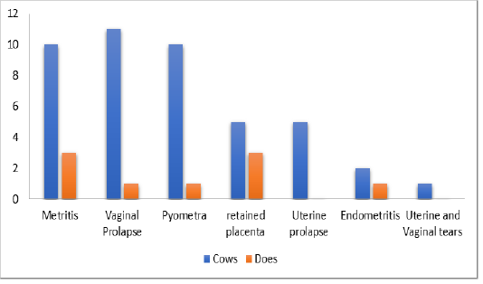
Figure 1: Incidence of postpartum disease and disorders in cows and does in the Klang Valley, Malaysia
Table 1: Distribution of recorded postpartum disease according to species in the Klang Valley, Malaysia
| Incidence of postpartum cases | Percentage | |
| Cattle | 44 | 83.0 |
| Goats | 9 | 17.0 |
| Total | 53 | 100 |
Table 2: Incidence of postpartum disease and disorders in cows and does based on age in the Klang Valley, Malaysia
| Species | Age | Incidence | Percentage |
| Cows | Young | 25 | 56.8 |
| Old | 19 | 43.2 | |
| Does |
Old Young |
6 3 |
66.6 33.4 |
| Total | 53 |
Table 3: Incidence of postpartum diseases and disorders based on parity in cows and does in Klang Valley, Malaysia
| Parity | Incidence | Percentage |
| First | 41 | 77.4 |
| Second | 9 | 17.0 |
| Third | 3 | 5.7 |
| Total | 53 | 100 |
Discussion
Postpartum metritis, vaginal prolapse and pyometra were found to be the frequently occurring diseases in postpartum cows and does in this investigation. These conditions have been frequently reported in cattle and goats from different parts of this world in varying proportions. The three most commonly documented postpartum disease in this investigation are common in livestock in many parts of the world (Sheldon et al., 2006, Sharma et al., 2017). The impact of the occurrence of these diseases and disorders are better understood in economic terms. Postpartum metritis, vaginal prolapse and pyometra are significant postpartum economic diseases in animals and causes high losses due to increased postpartum anestrus period and involuntary culling (Deori and Phookan, 2015).
Postpartum metritis is observed to be the most common postpartum disease in ruminants in the Klang valley during the time under review. Postpartum metritis usually develops when a fluid filled uterus having necrotic debris are invaded by bacteria. Therapy of postpartum metritis is achieved by using systemic antibiotics (Deori and Phookan, 2015). Other methods of managing post-partum metritis is to perform a uterine lavage with 0.9% NaCl. Uterine lavage is earlier reported to be the method of choice in the treatment of puerperal metritis in the Klang Valley (Abdullah et al., 2015a). Postpartum metritis is found to be the common postpartum disease in cows and goats in the Klang Valley between 2013 and 2017 in this current study. The occurrence of postpartum metritis in livestock have been reported to vary based on season and geographical location. The incidence of postpartum metritis in this investigation was found to be 24.5% which lies within reported cases in other parts of the world as incidence rates of 20-50% were reported in Canada and the US (Overton and Fetrow, 2008; Dubuc et al., 2010). In Europe, a lower postpartum metritis rate of 20-15% have been reported in livestock in Belgium (Opsomer et al., 2000). The incidence rate reported in this study is lower than the incidence rate of 28-39% as reported by Balasundaran et al. (2011) in India. Investigations into the occurrence of metritis in domestic ruminants has received considerable attention to date. Metritis reduces pregnancy rates during postpartum period and in subsequent reproductive performance in af-
Table 4: Chemotherapy of postpartum diseases in ruminants in Klang valley, Malaysia
| Class of drug/therapy used | Route | Drug used | Proportion | Percentage |
| Antibiotic | Intramuscular | Penicillin | 28 | 70 |
| Intramuscular | Tetracyclines | 12 |
30 |
|
| Anti-inflammatory | Intramuscular | Flunixin meglumine | 9 | 81.8 |
| Intramuscular | Dexamethasone | 2 |
18.2 |
|
| Uterine lavage | Intra uterine | 0.9% NaCl | 48 | 90.6 |
| Oxytetracyclines | 5 |
9.4 |
Table 5: Frequency of follow up of postpartum diseases and disorders in ruminants in the Klang Valley, Malaysia
| Proportion | Responses | Percentage |
| Yes | 25 | 47.2 |
| No | 28 | 52.8 |
| Total | 53 | 100 |
fected animals. It is a localised condition in the uterus and does not cause mortality in affected animals and there has been no association with loss of milk production affected animals. However economic losses are accrued through direct cost of treatment, examination, repeated inseminations and hormonal therapies. Furthermore, such cows spend some of their productive lifetime with such condition leading to fewer pregnancy rates and incidences of early culling.
Vaginal prolapse was the second most commonly occurring postpartum disease in ruminants in the Klang valley within the time under review. Breed predisposition, dietary and hormonal imbalances as well as hormonal imbalances are common risk factors for the occurrence of vaginal prolapse in animals (Miesner and Anderson, 2008). In a separate report, Abdullah et al. (2014b) reported that dietary imbalance was a leading cause of vaginal prolapse in a cow with a case of grade I vaginal prolapse. The incidence of vaginal prolapse observed in this investigation (22.6%) falls between the range of reported incidence from few document cases from other geographical locations. Incidence rates of 4.3% have been reported in livestock in Bangladesh (Sarder et al., 2015). Contrastingly, a higher incidence rate of 52.4% has equally been reported in India (Bhattacharyya et al., 2012). In this investigation, most cases of vaginal prolapse observed were Grade I and II prolapses based on the scale described by Miesner and Anderson (2009). Grade I and II vaginal prolapse are effectively managed using Buhner’s suture and such suture is usually placed around the lateral and ventral commissure of the vulva. This approach has been documented as the method of choice for management of vaginal prolapse within the study area (Abdullah et al., 2014b; 2015b; Yimer et al., 2016). Vaginal prolapse is common in cattle and quite rare in goat populations. The condition is usually seen around peri partum period in ruminants. Culling of animals affected suffering from vaginal prolapse is usually recommended due to the possibility of re-occurrence and ensuing economic losses in affected animals.
Pyometra was the third most common diseases among postpartum cows and does in the Klang Valley. This condition is characterised by the presence of an active corpus luteum together with the accumulation of uterine fluid with variable amounts of echo density. Postpartum pyometra is generally rare in animals and incidence rates has been placed at <5% (Sheldon et al., 2008). Similarly, Busch and Kuhnke (2000) reported a low incidence of 1.2% cases in cows in Germany with cases of pyometra. The incidence rate of postpartum pyometra (22.6%) in this investigation was found to be higher than previous reports. Although the cause of this variation was not investigated, this could be due to species variation or immune response of animals in different geographical locations. This condition is mainly caused by a persisting corpus luteum during postpartum Busch and Kuhnke (2000). This condition can have substantial economic importance as affected animals cannot conceive. Treatment of pyometra is achieved with injections of PGF2α and thereafter affected animals resume normal cyclicity.
Follow up visits to monitor the recovery rate and provide necessary additional therapy to animals is vital to restoration of normal body physiology and function. Follow up have been reported in some studies where disease outbreaks have been reported and where clinical management of diseases is performed (Haven et al., 1993; Cable et al., 2004; Meroc et al., 2015). The rate of follow up as observed in this study is low. This could be due to a rapid recovery following first therapy or due to culling and subsequent sale to reduce economic losses through feeding and continuous therapy. It could also be due to remote locations of the farms where follow ups may not be possible. Despite all these possibilities, follow up therapies must be an integral part of therapeutic approach in all diseases of animals.
This study reported that more postpartum diseases occurred in younger animals than in older animals. Similarly, more postpartum diseases were seen in animals during their first postpartum than in subsequent parturition. This however does not agree with earlier reports by Ayele et al. (2014) who in a similar study reported that postpartum disease is more common in older livestock. They also reported that the incidence of postpartum disease in animals is directly proportional to the frequency of parturition. Treatment of postpartum diseases and disorders are usually directed towards improving the fertility of the affected animals. This is because these conditions have significant influence on the productivity of affected animals. Treatment of metritis, pyometra, endometritis has successfully been achieved using antibiotics (Haimerl et al., 2017). Such antibiotics should be effective against the main uterine pathogens and should not inhibit normal defence mechanism of the animal. Penicillin and tetracyclines are frequently reported in literature for the management of bacterial infection during postpartum (Königsson et al., 2001; Deori and Phookan, 2015; El-Khadrawy et al., 2015; Lima, 2018). These drugs are widely used in the Klang Valley for the management of postpartum diseases as documented in farm records.
Conclusion
This study shows that many postpartum diseases occur in cows than does in the Klang Valley. Frequent postpartum diseases were metritis, vaginal prolapse pyometra and retained placenta. The study also showed that these diseases were more common in animals after their first parturition. Flunixin meglumine, oxytetracyclines and penicillin are the common drugs that were successfully used in managing postpartum diseases and disorders in the Klang Valley. However, veterinarians, animal health workers and animal owners need to collectively improve follow up treatments on animals having postpartum diseases and disorders until affected animals return to normal reproductive capacity.
Acknowledgement
The authors wish to acknowledge with thanks the owners of all the farms visited and for giving us access to their farm records.
Conflict of interest
None to declare.
Authors Contribution
All authors contributed equally and approved the final manuscript.
References


