Advances in Animal and Veterinary Sciences
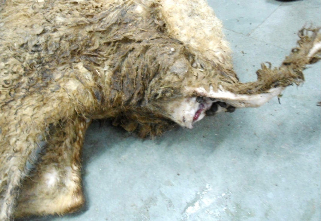
Hind-quarter of the animal is soiled with faeces due to diarrhoea, gaping of the anus and loss of lustre of hairs
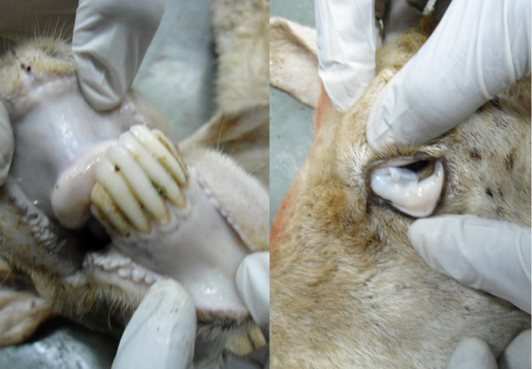
Buccal and conjunctival mucous membranes were pale and anaemic
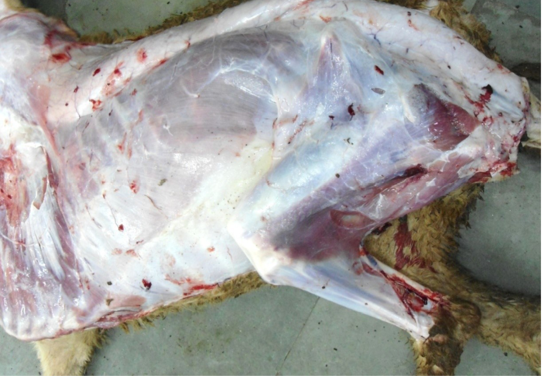
Carcass was pale in colour, hide bound condition, gelatinization of subcutaneous (SC) fat and SC fat was not in appropriate amount
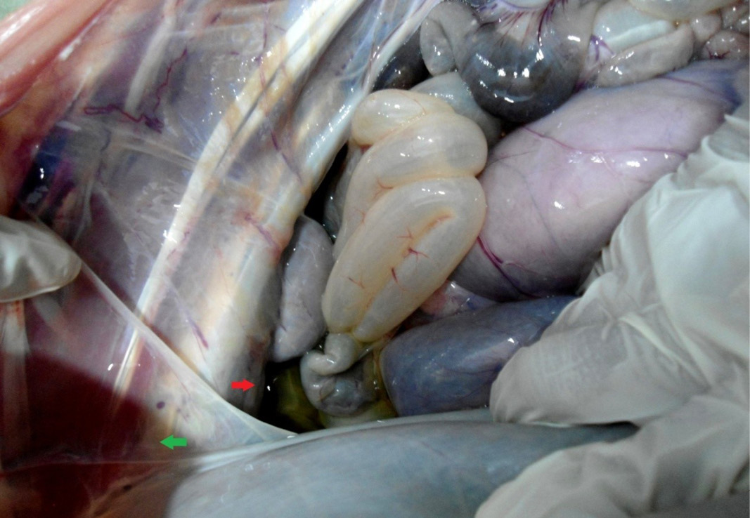
Hydrothorax (green arrow) and ascites (red arrow) due to hypoproteinemia
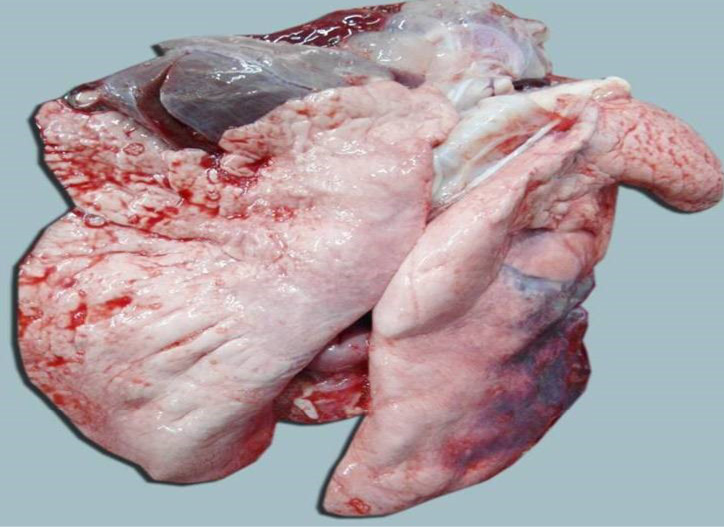
Lungs were pale and emphysematous and heart was pale and gelatinization of epicardial fat
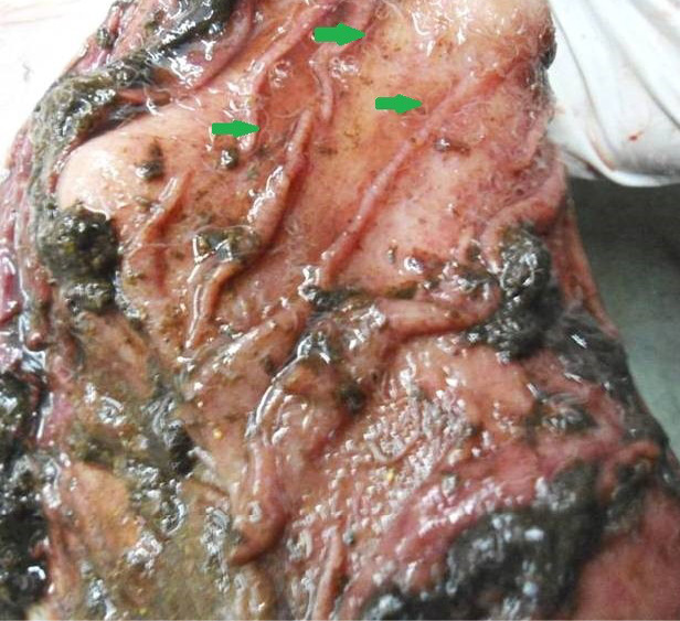
Abomasal mucosa was severely congested; pin point petechial haemorrhages and numerous minute hair like Haemonchus contortus worms (green arrows) were present
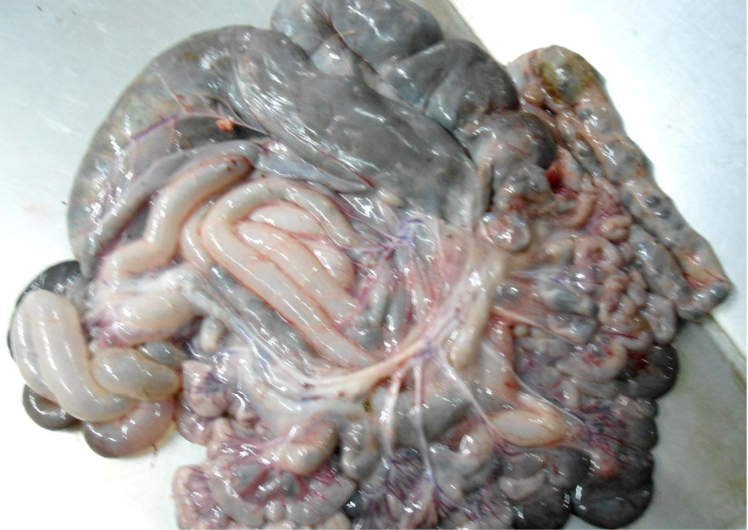
Intestine was pale in colour due to anaemia. Mesenteric lymph node was enlarged, edematous and pale in color
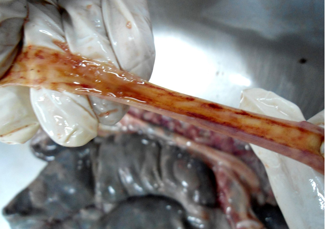
Small intestinal mucosa was congested, petechial to ecchymotic haemorrhages, thickened intestinal wall and watery intestinal contents
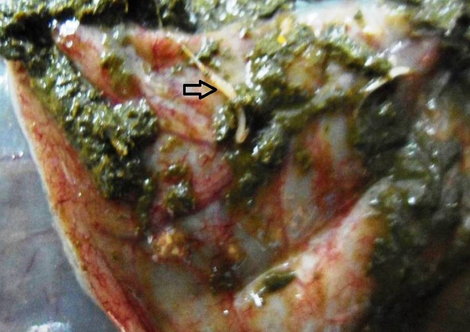
Caecal mucosa was congested; pin-point hemorrhages and few whipworms (arrow) were attached with mucosa
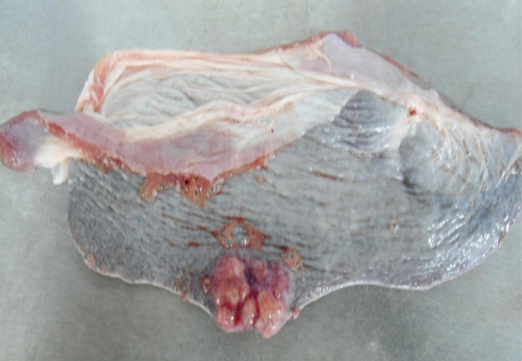
Multiple tiny cysts filled with straw yellow colour fluid were present in the left lateral border of the spleen
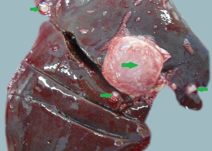
Liver was swollen with rounded borders, hard in consistency and multiple different size calcified masses (green arrows) were noticed
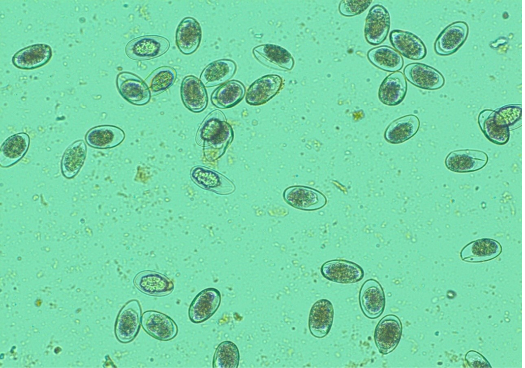
Numerous H. contortus eggs- thin-shelled, oval shape with equal poles, edges mombate and morula not fully filled the cavities of the eggs
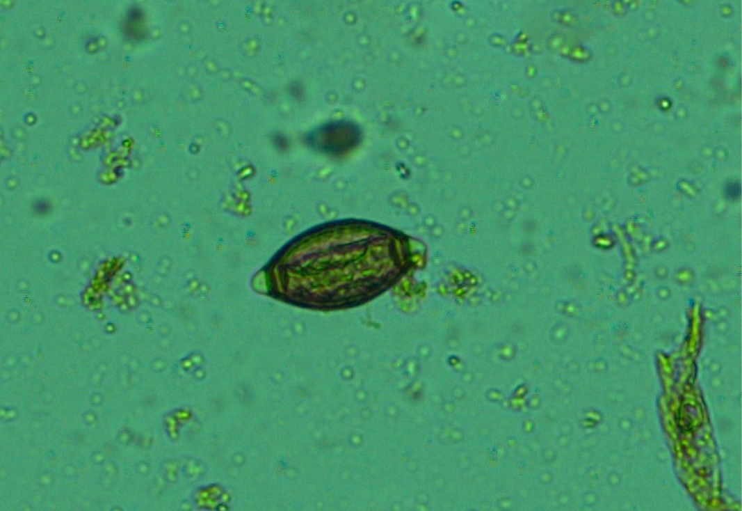
Whip worm (Trichuris ovis) eggs- dark brown colour, thick-walled, barrel shape with unsegmented embryo and transparent polar plugs at both ends
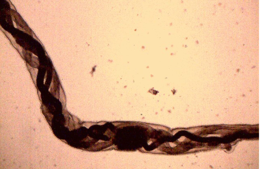
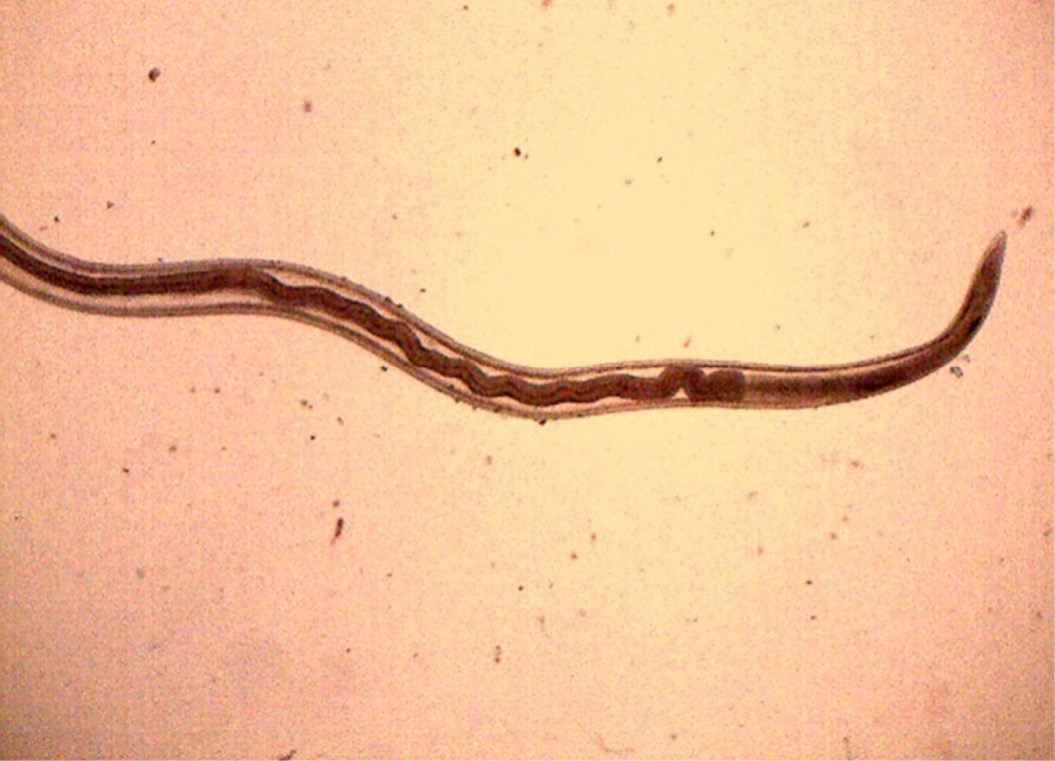
Female H. contortus worms- white uteri and ovaries winding around the red blood-filled intestine give a twisted or barber pole appearance
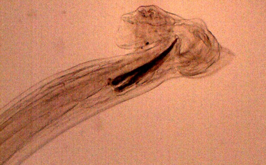
Anterior end of H. contortus was characterized by small funnel shaped buccal capsule and spine like pair of cervical papillae
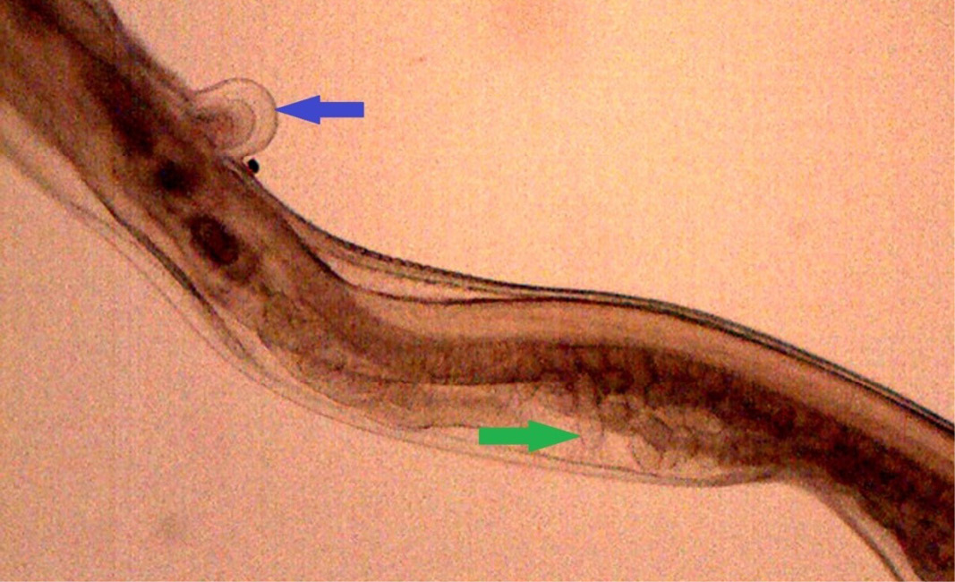
Posterior end of female H. contortus was characterized by knob type vulval flap (blue arrow) which covers the vulva and the uteri are fully packed with oval eggs (green arrow)
Posterior end of male H. contortus- copulatory bursa with well-developed asymmetrical dorsal lobes supported by inverted ‘Y’ shaped dorsal rays. Spicules are equal, short and end in knobs and are provided with barbs near the tip.
Abomasum showed haemorrhages, oedema, severe congestion of blood vessels in lamina propria and desquamation in the apical border of the villi
Prominent eosinophilic infiltration in the mucous and gastric glands of abomasum and infiltration of mononuclear cells especially lymphocytes
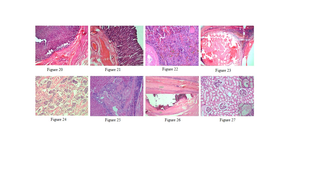
Figure 20: Thickening of abomasal mucosa due to hyperplasia of mucous and gastric glands
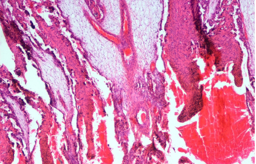
Figure 21: Small intestine showed highly congested capillaries in sub mucosa and thickened muscular layer
Figure 22: Spleen showed hemosiderosis (green arrows), depletion of red bulb area and prominent trabeculae
Figure 23: Spleen contains encapsulated cyst with homogenous eosinophilic material and cyst is lined by thick fibrous wall
Figure 24: Prescapular lymph node showed severe depletion of lymphocyte in both cortical and medullary region and prominent trabeculae
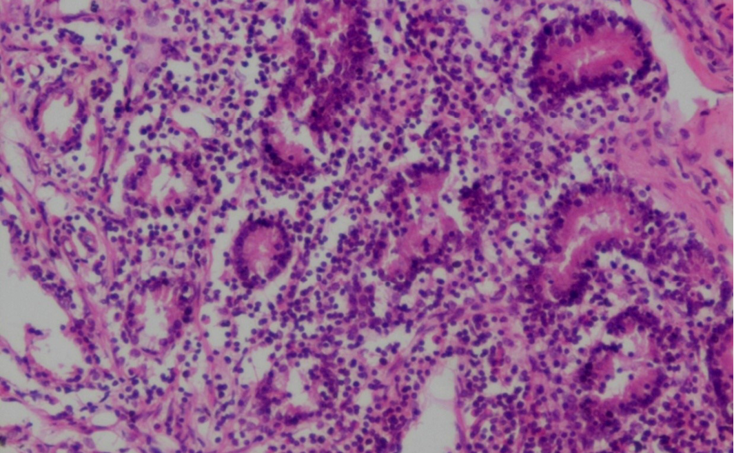
Figure 25: Liver showed mild bile duct hyperplasia, narrowing of bile duct lumen, degeneration of hepatocyte and mononuclear cell infiltration especially lymphocytes
Figure 26: Liver showed homogenous basophilic calcified mass (black arrows) which lined by thick fibrous capsule (green arrows)
Figure 27: Glomerulus showed eosinophilic protenacious edematous fluid and mild degenerative changes in the epithelium. Inset showing atrophy of the glomeruli


